. 2004 Jun;121(6):493-9.
doi: 10.1007/s00418-004-0653-5. Epub 2004 May 15.

Mepe is expressed during skeletal development and regeneration

Affiliations

Mepe is expressed during skeletal development and regeneration

Chuanyong Lu et al. Histochem Cell Biol. 2004 Jun.

Abstract

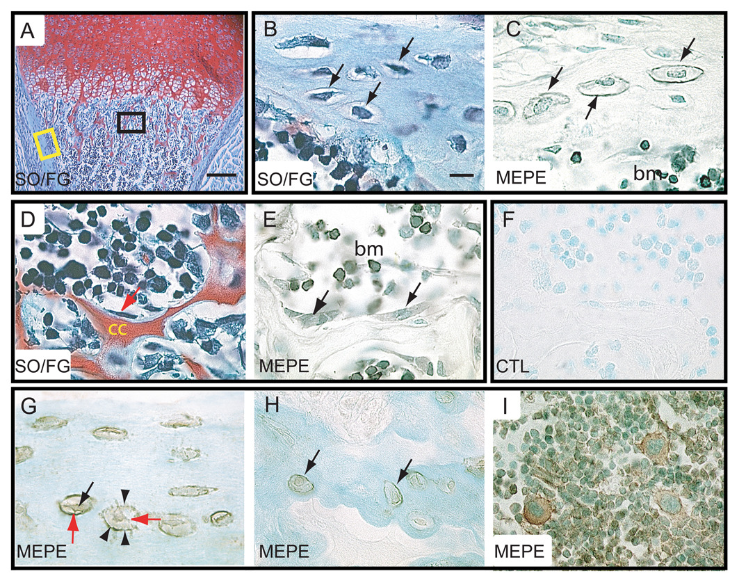
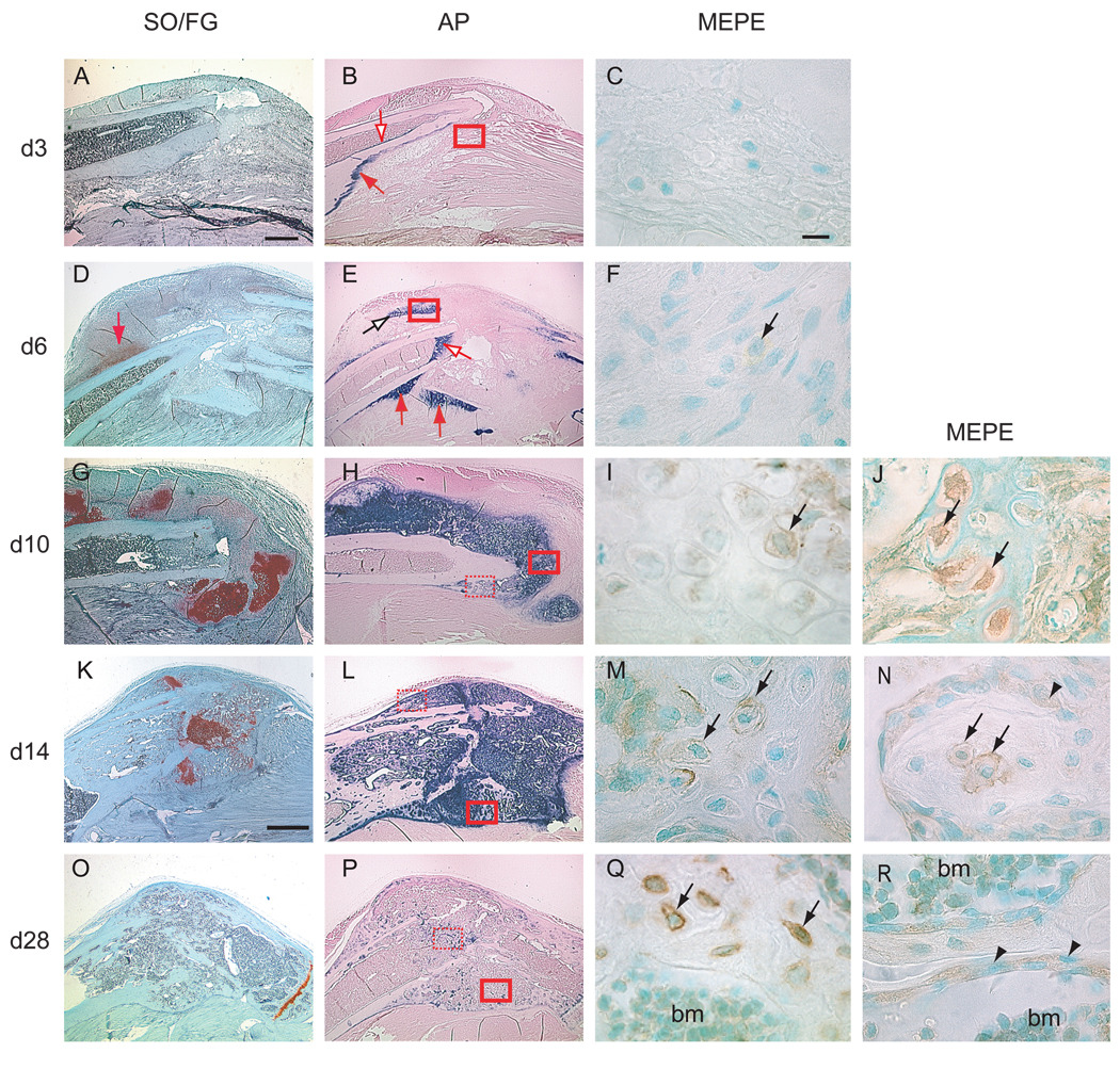
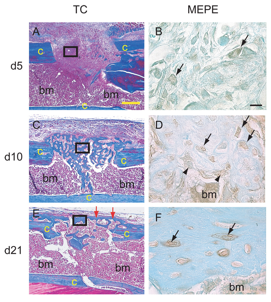
a bone metabolism regulator that is expressed by osteocytes in normal adult bone. Here, we used an immunohistochemical approach to study whether Mepe has a role in murine long bone development and regeneration. Our data showed that Mepe protein was produced by osteoblasts and osteocytes during skeletogenesis, as early as 2 days postnatal. During the healing of non-stabilized tibial fractures, which occurs through endochondral ossification, Mepe expression was first detected in fibroblast-like cells within the callus by 6 days postfracture. By 10 and 14 days postfracture (the hard callus phase of repair), Mepe was expressed within late hypertrophic chondrocytes and osteocytes in the regenerating tissues. Mepe became externalized in osteocyte lacunae during this period. By 28 days postfracture (the remodeling phase of repair), Mepe continued to be robustly expressed in osteocytes of the regenerating bone. We compared the Mepe expression profile with that of alkaline phosphatase, a marker of bone mineralization. We found that both Mepe and alkaline phosphatase increased during the hard callus phase of repair. In the remodeling phase of repair, Mepe expression levels remained high while alkaline phosphatase activity decreased. We also examined Mepe expression during cortical bone defect healing, which occurs through intramembranous ossification. Mepe immunostaining was found within fibroblast-like cells, osteoblasts, and osteocytes in the regenerating bone, through 5 to 21 days postsurgery. Thus, Mepe appears to play a role in both long bone regeneration and the latter stages of skeletogenesis.


Figures

Figure 1
Figure 1
Immunolocalization of MEPE protein in developing and mature long bones. (A–F) Longitudinal sections through the humerus of a 2-day post-natal mouse. (A) SO/FG staining showed hypertrophic cartilage in the process of being replaced by trabecular bone. (B) Higher magnification of the cortex showed osteocytes (arrows) embedded in bone matrix (corresponding to yellow box in A). (C) MEPE immunostaining on an adjacent section was localized in osteocytes (arrows) of the cortex, along osteocyte lacunae (arrowheads), and in bone marrow cells. (D) Higher magnification in the metaphysis (corresponding to the black box in A) showed osteoblasts (arrow) lining the remnants of calcified cartilage (cc) matrix. (E) MEPE immunostaining on an adjacent section was detected in both osteoblasts (arrows) and bone marrow cells. (F) Negative control showed the lack of MEPE staining. (G–I) Longitudinal section through a 12-week old mouse humerus. (G) Osteocytes within cortical bone were positive for MEPE immunostaining. MEPE immunoreactivity was found intracellularly (black arrow) and on the wall of osteocyte lacunae (arrowheads). MEPE immunoreactivity was also detected in the cell processes (red arrows). (H) Osteocytes in trabecular bone were also positive for MEPE immunostaining. (I) MEPE immunostaining in bone marrow cells. bm= bone marrow, cc = calcified cartilage, SO/FG = Safranin O/Fast Green staining, CTL= control. Scale bar in A = 500 µm, B–I = 10 µm.
Figure 2
Figure 2
Distribution of MEPE protein in the callus tissue during non-stabilized fracture healing. (A, D, G, K, O) Safranin O/Fast Green staining on longitudinal sections through the fracture callus between 3 (d3) and 28 days (d28) post-fracture. Cartilage stained red. (B, E, H, L, P) Alkaline phosphatase (AP) staining (blue staining) on adjacent sections. (C, F, I, M, Q) MEPE immunostaining in area corresponding to solid red boxes in (B, E, H, L, P) respectively. (J, N, R) MEPE immunostaining in area corresponding to the dashed red boxes in (H, L, P) respectively. (A) At 3 days post-fracture, SO/FG staining indicated no proteoglycan accumulation, as shown by the lack of red staining. (B) Minimal AP activity was evident on the endosteum (empty arrow) and periosteum (red arrow). (C) No MEPE immunostaining was detectable around the fracture site. (D) By 6 days, cartilage islands within the callus were visible by SO/FG staining (red arrow). (E) AP activity was seen around these cartilage islands (empty black arrow), in the periosteum (red arrows), and in the endosteum (empty red arrow). (F) Weak MEPE immunostaining was found in some of the cells within these cartilage islands (arrow). (G) By 10 days, abundant cartilage was present in the fracture callus. (H) AP activity was strong throughout the cartilage callus and in the new periosteal bone. (I) MEPE immunostaining was detectable in late hypertrophic chondrocytes within the callus (arrow). (J) MEPE immunostaining was detected in osteocytes (arrows) embedded in the new bone. MEPE immunostaining was associated with the cell body and the cell processes. (K) By 14 days, the amount of cartilage within the callus was diminished. (L) AP activity was evident throughout the callus. (M) MEPE immunostaining was found in the pericellular matrix of late hypertrophic chondrocytes (arrows) at the ossification front. (N) MEPE immunostaining was localized in the osteocytes and in the lacunae (arrows) within the newly formed trabecular bone. Osteoblasts (arrowheads) lining the trabecular bone exhibited weak MEPE immunostaining. (O) By 28 days, the hypertrophic cartilage was completely resorbed and (P) AP activity was decreased. (Q) Strong MEPE immunostaining was seen in osteocytes in the new bone (arrows). (R) Osteoblasts (arrowheads) adjacent to trabecular bone and bone marrow cells (bm) were also positive for MEPE immunostaining. bm = bone marrow. Scale bars: A, B, D, E, G, H = 0.5mm. K, L, O, P = 1mm. MEPE = 10 µm.
Figure 3
Figure 3
Immunodetection of MEPE protein during healing of a cortical bone defect. (A, C, E) Longitudinal sections through a 1mm cortical bone defect stained with Trichrome. (B, D, F) MEPE immunostaining on adjacent sections corresponding to the area of solid black boxes in (A,C, E) respectively. (A) By 5 days post-surgery, granulation tissue filled the defect. (B) Weak MEPE immunostaining was detected in fibroblastic cells within the defects (arrows). (C) By 10 days, new bone had formed in the cortical defect. (D) Osteocytes (arrows) and osteoblasts (arrowheads) in the new bone exhibited MEPE immunostaining. (E) By 21 days, new bone within the cortical defect was remodeled and a new cortex had formed (arrows). (F) MEPE immunostaining was strong in the osteocytes and their pericellular matrix within the new cortex (arrows). Bone marrow cells were also positive for MEPE immunostaining. c = cortex, bm = bone marrow, TC = Trichrome staining. Scale bars: A, C, E = 300 µm, B, D, F = 10 µm.

Similar articles

Cited by

References

    1. Albrecht UEG, Helms JA, Lin H. Visualization of gene expression patterns by in situ hybridization. In: Daston GP, editor. Molecular and cellular methods in developmental toxicology. Boca Raton, FL: CRC Press; 1997. pp. 23–48.
    1. Argiro L, Desbarats M, Glorieux FH, Ecarot B. Mepe, the gene encoding a tumor-secreted protein in oncogenic hypophosphatemic osteomalacia, is expressed in bone. Genomics. 2001;74:342–351. - PubMed
    1. Boden SD, Kaplan FS. Calcium homeostasis. Orthop Clin North Am. 1990;21:31–42. - PubMed
    1. Colnot C, Thompson Z, Miclau T, Werb Z, Helms JA. Altered fracture repair in the absence of MMP9. Development. 2003;130:4123–4133. - PMC - PubMed
    1. Doppelt SH. Vitamin D, rickets, and osteomalacia. Orthop Clin North Am. 1984;15:671–686. - PubMed

Publication types

MeSH terms

Substances

LinkOut - more resources