

Relationship between encephalopathy and portal vein-vena cava shunt: value of computed tomography during arterial portography
- PMID: 15222041
- PMCID: PMC4572235
- DOI: 10.3748/wjg.v10.i13.1939
Relationship between encephalopathy and portal vein-vena cava shunt: value of computed tomography during arterial portography
Abstract
Aim: To assess the value of computed tomography during arterial portography (CTAP) in portal vein-vena cava shunt, and analysis of the episode risk in encephalopathy.
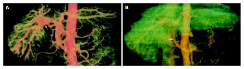
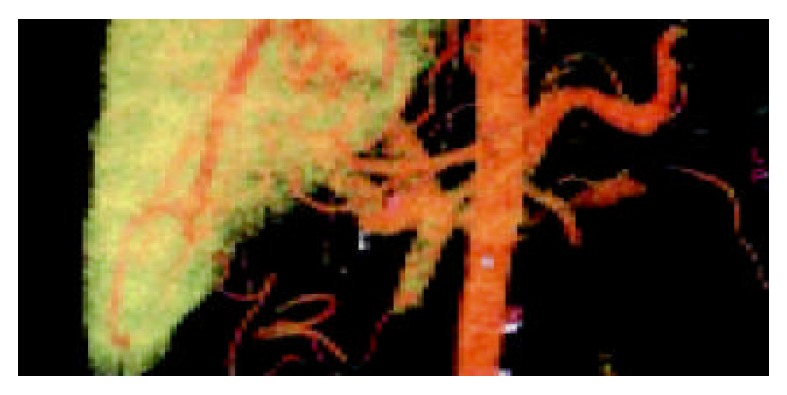
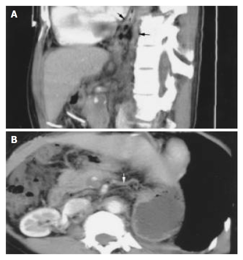
Methods: Twenty-nine patients with portal-systemic encephalopathy due to portal hypertension were classified by West Haven method into grade I(29 cases), grade II(16 cases), grade III(10 cases), grade IV( 4 cases). All the patients were scanned by spiral-CT. Plane scans, artery phase and portal vein phase enhancement scans were performed, and the source images were thinly reconstructed to 1.25 mm. We reconstructed the celiac trunk, portal vein, inferior vena cava and their branches and subjected them to three-dimensional vessel analysis by volume rendering (VR) technique and multiplanar volume reconstruction (MPVR) technique. The blood vessel reconstruction technique was used to evaluate the scope and extent of portal vein-vena cava shunt, portal vein emboli and the fistula of hepatic artery-portal vein. The relationship between the episode risk of portal-systemic encephalopathy and the scope and extent of portal vein-vena cava shunt, portal vein emboli and fistula of hepatic artery- portal vein was studied.
Results: The three-dimensional vessel reconstruction technique of spiral-CT could display celiac trunk, portal vein, inferior vena cava and their branches at any planes and angles and the scope and extent of portal vein-vena cava shunt, portal vein emboli and the fistula of hepatic artery- portal vein. In twenty-nine patients with portal-systemic encephalopathy, grade I accounted for 89.7% esophageal varices, 86.2% paragastric varices; grade II accounted for 68.75% cirsomphalos, 56.25% paraesophageal varices, 62.5% retroperitoneal varices and 81.25% dilated azygos vein; grade III accounted for 80% cirsomphalos, 60% paraesophageal varices, 70% retroperitoneal varices, 90% dilated azygos vein, and part of the patients in grades II and III had portal vein emboli and fistula of hepatic artery-portal vein; grade IV accounted for 75% dilated left renal vein, 50% paragallbladder varices, all the patients had fistula of hepatic artery- portal vein.
Conclusion: The three-dimensional vessel reconstruction technique of spiral-CT can clearly display celiac trunk, portal vein, inferior vena cava and their branches at any planes and angles and the scope and extent of portal vein-vena cava shunt. The technique is valuable for evaluating the episode risk in portal-systemic encephalopathy.
Figures

Similar articles
-
Diagnostic value of 16 slices spiral-CT for portal vein disorders.J Huazhong Univ Sci Technolog Med Sci. 2004;24(3):300-2. doi: 10.1007/BF02832020. J Huazhong Univ Sci Technolog Med Sci. 2004. PMID: 15315356
-
[The value of multi-slice spiral computed tomography portography in assessing severity of liver cirrhosis and predicting episode risks of hepatic encephalopathy].Zhonghua Gan Zang Bing Za Zhi. 2014 Jul;22(7):509-13. doi: 10.3760/cma.j.issn.1007-3418.2014.07.007. Zhonghua Gan Zang Bing Za Zhi. 2014. PMID: 25203802 Chinese.
-
Three-dimensional CT demonstration of intrahepatic portosystemic venous shunt draining into the inferior vena cava.Br J Radiol. 1997 Apr;70(832):418-20. doi: 10.1259/bjr.70.832.9166081. Br J Radiol. 1997. PMID: 9166081
-
Angiography in portal hypertension.Gastroenterol Clin North Am. 1992 Mar;21(1):61-83. Gastroenterol Clin North Am. 1992. PMID: 1568778 Review.
-
Portal-systemic encephalopathy in non-cirrhotic patients: classification of clinical types, diagnosis and treatment.J Gastroenterol Hepatol. 2000 Sep;15(9):969-79. doi: 10.1046/j.1440-1746.2000.02283.x. J Gastroenterol Hepatol. 2000. PMID: 11059925 Review.
Cited by
-
Radiological score for hemorrhage in the patients with portal hypertension.Int J Clin Exp Pathol. 2015 Sep 1;8(9):11517-23. eCollection 2015. Int J Clin Exp Pathol. 2015. PMID: 26617884 Free PMC article.
References
-
- Butterworth RF. Hepatic encephalopathy: a neuropsychiatric disorder involving multiple neurotransmitter systems. Curr Opin Neurol. 2000;13:721–727. - PubMed
-
- Calculli L, Casadei R, Amore B, Albini Riccioli L, Minni F, Caputo M, Marrano D, Gavelli G. The usefulness of spiral Computed Tomography and colour-Doppler ultrasonography to predict portal-mesenteric trunk involvement in pancreatic cancer. Radiol Med. 2002;104:307–315. - PubMed
-
- Ferenci P, Lockwood A, Mullen K, Tarter R, Weissenborn K, Blei AT. Hepatic encephalopathy--definition, nomenclature, diagnosis, and quantification: final report of the working party at the 11th World Congresses of Gastroenterology, Vienna, 1998. Hepatology. 2002;35:716–721. - PubMed
-
- Francis IR, Cohan RH, McNulty NJ, Platt JF, Korobkin M, Gebremariam A, Ragupathi KI. Multidetector CT of the liver and hepatic neoplasms: effect of multiphasic imaging on tumor conspicuity and vascular enhancement. AJR Am J Roentgenol. 2003;180:1217–1224. - PubMed
-
- Matoba M, Yokota H, Yabuno K, Kuga G, Tinami H, Yamamoto I. [Evaluation of two injection protocols by time-density curves for possible application to hepatic dynamic and upper abdominal CT angiography in MDCT: high concentration (350 mgI/ml) with conventional volume (100 ml) vs. conventional concentration (300 mgI/ml) with larger volume (150 ml) and higher injection rate] Nihon Igaku Hoshasen Gakkai Zasshi. 2003;63:98–102. - PubMed
Publication types
MeSH terms
LinkOut - more resources
Full Text Sources
Medical
