

Role of curved planar reformations using multidetector spiral CT in diagnosis of pancreatic and peripancreatic diseases
- PMID: 15222042
- PMCID: PMC4572236
- DOI: 10.3748/wjg.v10.i13.1943
Role of curved planar reformations using multidetector spiral CT in diagnosis of pancreatic and peripancreatic diseases
Abstract
Aim: To investigate the role of curved planar reformations using multidetector spiral CT (MSCT) in diagnosis of pancreatic and peripancreatic diseases.
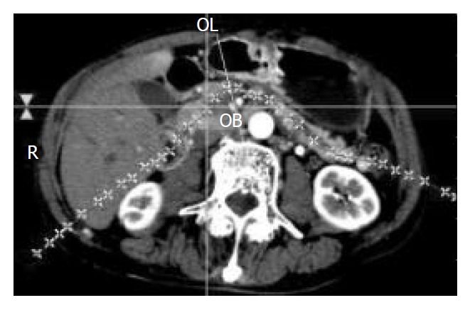
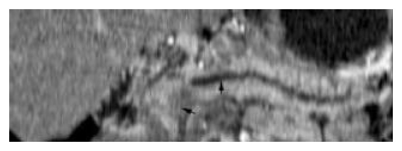
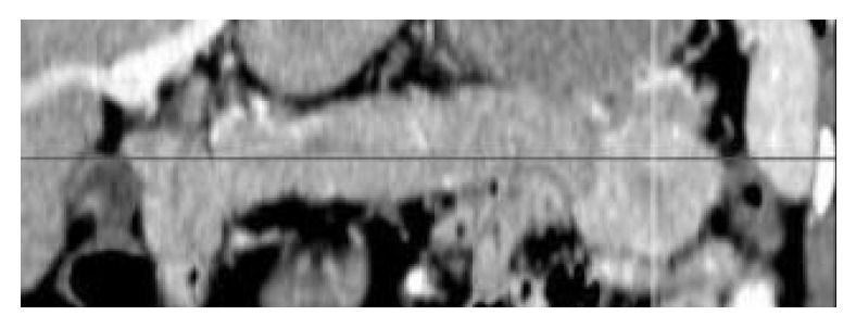
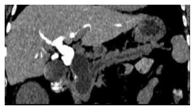
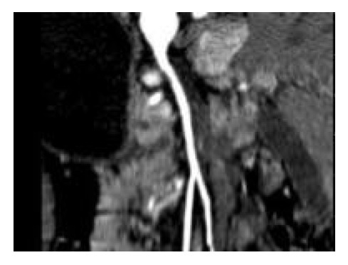
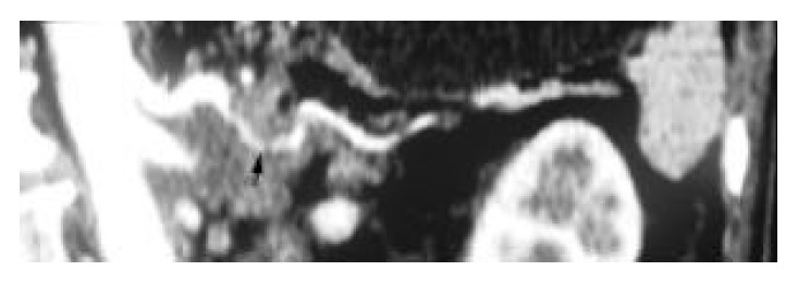
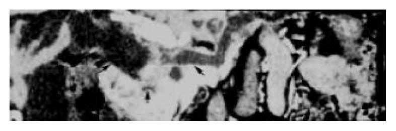
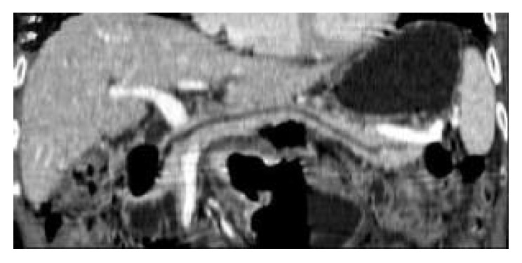
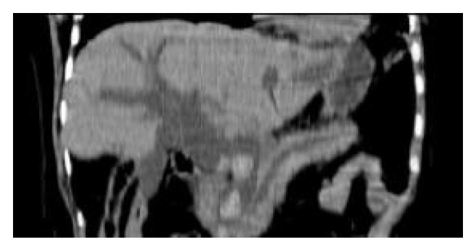
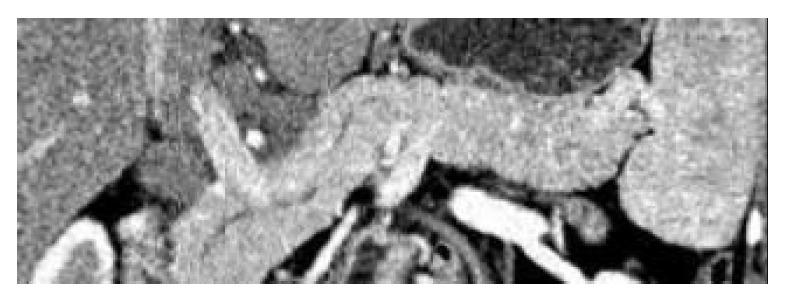
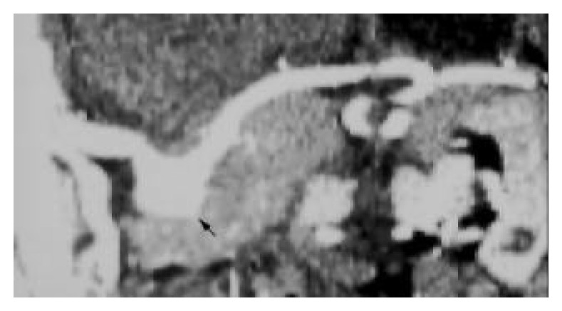
Methods: From October 2001 to September 2003, 47 consecutive patients with pancreatic or peripancreatic diseases, which were confirmed by operation, endoscopic retrograde cholangiopancreatography and clinical follow-up, were enrolled in this study. CT scanning was performed at a MSCT with four rows of detector. A set of images with an effective thickness of 1.0-2.0 mm and a gap of 0.5-1.0 mm (50% overlap) were acquired in all patients for post-processing. Curved planar reformations were carried out by drawing a curved line on transverse source images, coronal or sagittal multiplanar reformations according to certain anatomic structures (such as cholangiopancreatic ducts or peripancreatic vessels) and the position of lesion.
Results: With thin collimation, MSCT could acquire high-quality curved planar reformations to display the profile of the whole pancreas, to trace the cholangiopancreatic ducts and peripancreatic vessels, and to show the relationship of lesions with pancreas and peripancreatic anatomic structures in one curved plane, which facilitates diagnosis and rapid communication of diagnostic information with referring physicians.
Conclusion: MSCT with thin collimation could be used to create high-quality curved planar reformations in evaluating pancreatic and peripancreatic diseases with pertinent anatomic information and relative pathologic signs to facilitate the diagnosis and enhance communication with the referring physician. Curved planar reformations can serve as supplements for transverse images in diagnosis and management of pancreatic and peripancreatic diseases.
Figures
Similar articles
-
Local staging of pancreatic carcinoma with multi-detector row CT: use of curved planar reformations initial experience.Radiology. 2002 Dec;225(3):759-65. doi: 10.1148/radiol.2253010886. Radiology. 2002. PMID: 12461258
-
[Curved planar reformations in multi-slice spiral CT in pancreatic adenocarcinoma: prediction of invasion of pancreatic and peripancreatic ductal structures].Zhongguo Yi Xue Ke Xue Yuan Xue Bao. 2006 Feb;28(1):71-5. Zhongguo Yi Xue Ke Xue Yuan Xue Bao. 2006. PMID: 16548194 Chinese.
-
Multidetector-row CT and volumetric imaging of pancreatic neoplasms.Gastroenterol Clin North Am. 2002 Sep;31(3):881-96. doi: 10.1016/s0889-8553(02)00029-8. Gastroenterol Clin North Am. 2002. PMID: 12481736 Review.
-
Peripancreatic arteries in thin-section multislice helical CT.Abdom Imaging. 2001 May-Jun;26(3):234-42. doi: 10.1007/s002610000165. Abdom Imaging. 2001. PMID: 11429946
-
Pancreatic and peripancreatic diseases mimicking primary pancreatic neoplasia.Radiographics. 2005 Jul-Aug;25(4):949-65. doi: 10.1148/rg.254045167. Radiographics. 2005. PMID: 16009817 Review.
Cited by
-
Evaluating blunt pancreatic trauma at whole body CT: current practices and future directions.Emerg Radiol. 2013 Dec;20(6):517-27. doi: 10.1007/s10140-013-1133-9. Epub 2013 Jun 6. Emerg Radiol. 2013. PMID: 23739797 Review.
-
Multi-detector computed tomography of acute abdomen.Eur Radiol. 2005 Dec;15(12):2435-47. doi: 10.1007/s00330-005-2897-4. Epub 2005 Aug 27. Eur Radiol. 2005. PMID: 16132914 Review.
-
Portal Vein/Aorta Ratio in Dogs with Acquired Portosystemic Collaterals.J Vet Intern Med. 2017 Sep;31(5):1382-1387. doi: 10.1111/jvim.14802. Epub 2017 Aug 14. J Vet Intern Med. 2017. PMID: 28804949 Free PMC article.
-
Validation of a convolutional neural network for the automated creation of curved planar reconstruction images along the main pancreatic duct.Jpn J Radiol. 2023 Feb;41(2):228-234. doi: 10.1007/s11604-022-01339-1. Epub 2022 Sep 19. Jpn J Radiol. 2023. PMID: 36121623 Free PMC article.
-
[Multidetector computed tomography in abdominal emergencies].Radiologe. 2009 Jun;49(6):523-32. doi: 10.1007/s00117-008-1809-4. Radiologe. 2009. PMID: 19557461 Review. German.
References
-
- Rubin GD, Dake MD, Semba CP. Current status of three-dimensional spiral CT scanning for imaging the vasculature. Radiol Clin North Am. 1995;33:51–70. - PubMed
-
- Achenbach S, Moshage W, Ropers D, Bachmann K. Curved multiplanar reconstructions for the evaluation of contrast-enhanced electron beam CT of the coronary arteries. AJR Am J Roentgenol. 1998;170:895–899. - PubMed
-
- Portugaller HR, Schoellnast H, Tauss J, Tiesenhausen K, Hausegger KA. Semitransparent volume-rendering CT angiography for lesion display in aortoiliac arteriosclerotic disease. J Vasc Interv Radiol. 2003;14:1023–1030. - PubMed
-
- Takase K, Sawamura Y, Igarashi K, Chiba Y, Haga K, Saito H, Takahashi S. Demonstration of the artery of Adamkiewicz at multi- detector row helical CT. Radiology. 2002;223:39–45. - PubMed
Publication types
MeSH terms
LinkOut - more resources
Full Text Sources
Medical
Miscellaneous
