. 2004 Jul 5;200(1):35-46.
doi: 10.1084/jem.20040392. Epub 2004 Jun 28.

A new family of potent AB(5) cytotoxins produced by Shiga toxigenic Escherichia coli

Affiliations

A new family of potent AB(5) cytotoxins produced by Shiga toxigenic Escherichia coli

Adrienne W Paton et al. J Exp Med. .

Erratum in

  • J Exp Med. 2004 Dec 6;200(11):1525

Abstract

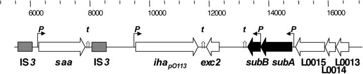
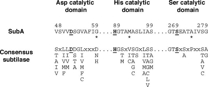
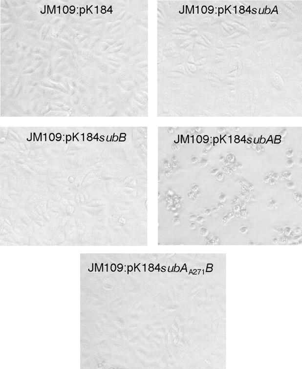
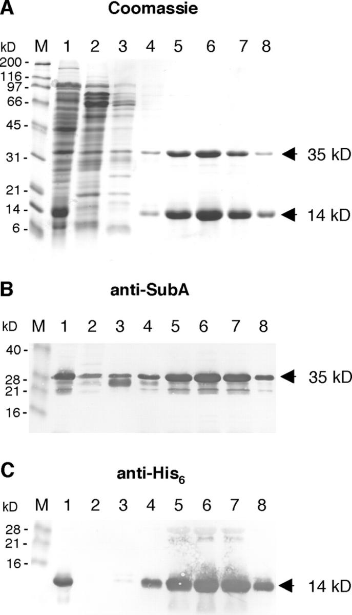
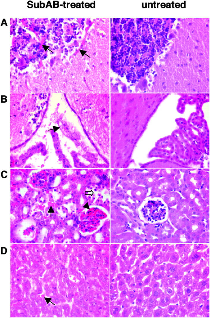
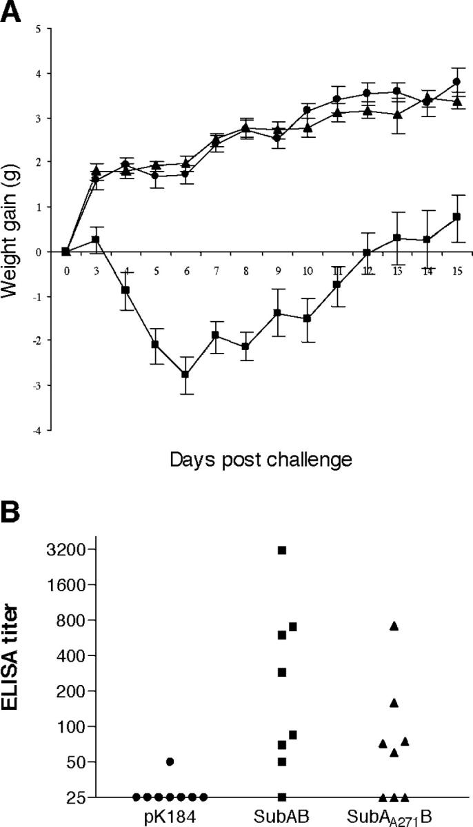
The Shiga toxigenic Escherichia coli (STEC) O113:H21 strain 98NK2, which was responsible for an outbreak of hemolytic uremic syndrome, secretes a highly potent and lethal subtilase cytotoxin that is unrelated to any bacterial toxin described to date. It is the prototype of a new family of AB(5) toxins, comprising a single 35-kilodalton (kD) A subunit and a pentamer of 13-kD B subunits. The A subunit is a subtilase-like serine protease distantly related to the BA_2875 gene product of Bacillus anthracis. The B subunit is related to a putative exported protein from Yersinia pestis, and binds to a mimic of the ganglioside GM2. Subtilase cytotoxin is encoded by two closely linked, cotranscribed genes (subA and subB), which, in strain 98NK2, are located on a large, conjugative virulence plasmid. Homologues of the genes are present in 32 out of 68 other STEC strains tested. Intraperitoneal injection of purified subtilase cytotoxin was fatal for mice and resulted in extensive microvascular thrombosis, as well as necrosis in the brain, kidneys, and liver. Oral challenge of mice with E. coli K-12-expressing cloned subA and subB resulted in dramatic weight loss. These findings suggest that the toxin may contribute to the pathogenesis of human disease.


Figures

Figure 1.
Figure 1.
Map of part of the megaplasmid pO113 from 98NK2. (top) The scale indicates the corresponding nt numbers in AF399919.3. The locations of the subA and subB ORFs are shown in black arrows; other ORFs are indicated by white arrows. The gray boxes represent incomplete IS3-like elements. The locations of putative promoters (P) and transcription terminator sequences (t) are also indicated.
Figure 2.
Figure 2.
Alignment of the three putative catalytic domains of SubA with the consensus sequence for Asp, His, and Ser catalytic domains of members of the subtilase family (reference 17). The numbers above the SubA fragments indicate the residue number of the terminal aas. Alternative consensus residues at a given position are shown vertically. The known active site residues in each subtilase catalytic domain (reference 17) are shown in bold type and underlined. *, SubA residues that do not match the consensus sequence.
Figure 3.
Figure 3.
Cytotoxicity of SubAB for Vero cells. Monolayers were treated with 1:80 dilutions of culture supernatant from the indicated strains for 72 h and photographed under phase-contrast microscopy.
Figure 4.
Figure 4.
Western blot analysis of subclones. Culture lysates of E. coli JM109 carrying the indicated plasmids were separated by SDS-PAGE, electroblotted onto nitrocellulose, and probed with anti-SubA (top) or anti-SubB (bottom). The Marker track contains BenchMark prestained protein markers (Invitrogen), and the approximate sizes of visible bands are indicated on the left side of the figure. The approximate sizes of the immunoreactive species are also indicated on the right.
Figure 5.
Figure 5.
Copurification of SubA and SubB. Crude lysate of E. coli Tuner™(DE3):pET-23(+)subAB was applied to a Ni-NTA column, washed, and eluted with a linear 0–500-mM imidazole gradient. 10 μl aliquots of the original lysate (lane 1) and fractions 3–9 (lanes 2–8) were separated by SDS-PAGE and stained with Coomassie blue (A). Lane M contains SDS-PAGE molecular weight standards (BioRad Laboratories), and the approximate sizes are indicated on the left of the figure. Alternatively, proteins were electroblotted onto nitrocellulose and probed with polyclonal anti-SubA (B) or monoclonal anti-His6 (C). Lane M contains BenchMark prestained protein markers (Invitrogen), and the approximate sizes of visible bands are indicated on the left side of the figure.
Figure 6.
Figure 6.
Immunofluorescent analysis. Vero cells were treated with purified SubAB for 48 h, fixed, permeabilized (except where indicated), and stained with mouse anti-SubA, anti-SubB, or nonimmune serum, followed by goat anti–mouse IgG-ALX488 conjugate. (A and C) Anti-SubA; (B and D) anti-SubB; (E) anti-SubA (nonpermeabilized); (F) anti-SubB (nonpermeabilized); (G) nonimmune serum; (H) nonimmune serum (nonpermeabilized); (I) anti-SubA without SubAB treatment; and (J) anti-SubB without SubAB treatment.
Figure 7.
Figure 7.
Histological examination of SubAB-treated mice. Mice were injected intraperitoneally with or without 1 μg of purified SubAB. After 6 d, the moribund toxin-treated mouse and the healthy control mouse were killed; brain, liver, and kidneys were removed; and fixed sections were examined histologically after staining with hematoxylin and eosin using a 40× objective. (A) Cerebellar cortex, showing hemolytic thrombi in molecular and granular layers from the toxin-treated mouse (arrows). (B) Choroid plexus, showing necrotic toxin-treated epithelial cells (arrow). (C) Renal cortex, showing interstitial thrombi (arrow), tubular necrosis (white arrow), and glomerular hyperemia (arrowhead) in the toxin-treated mouse. (D) Liver parenchyma, showing thrombi (arrow) and generalized hepatocyte necrosis in toxin-treated mouse.
Figure 8.
Figure 8.
Oral challenge of mice with SubAB-producing clones. Groups of streptomycin-treated BALB/C mice were fed ∼108 CFU of E. coli DH5αSR carrying pK184 (circles), pK184subAB (squares), or pK184subA A271 B (triangles). Drinking water was supplemented with 5 mg/ml streptomycin and 100 μg/ml kanamycin. (A) Effect on body weight. Individually identified mice were weighed on day 0 and daily from day 3. Data are mean weight gain (± SE), relative to weight of the respective mouse on day 0. The mean weights of mice in the three groups on day 0 were 18.7, 17.8, and 18.0 g, respectively. (B) Serum anti-SubAB levels. Sera collected on day 15 were assayed for antibodies to SubAB by ELISA. The minimum detectable titer was 50, and sera below this level have been assigned a nominal titer of 25.

References

    1. Fan, E., E.A. Merritt, C.L.M.J. Verlinde, and W.G.J. Hol. 2000. AB(5) toxins: structures and inhibitor design. Curr. Opin. Struct. Biol. 10:680–686. - PubMed
    1. Karmali, M.A. 1989. Infection by verocytotoxin-producing E. coli. Clin. Microbiol. Rev. 2:15–38. - PMC - PubMed
    1. Paton, J.C., and A.W. Paton. 1998. Pathogenesis and diagnosis of Shiga toxin-producing Escherichia coli infections. Clin. Microbiol. Rev. 11:450–479. - PMC - PubMed
    1. Schmidt, H., J. Scheef, H.I. Huppertz, M. Frosch, and H. Karch. 1999. Escherichia coli O157:H7 and O157:H(-) strains that do not produce Shiga toxin: phenotypic and genetic characterization of isolates associated with diarrhea and hemolytic-uremic syndrome. J. Clin. Microbiol. 37:3491–3496. - PMC - PubMed
    1. Paton, A.W., M.C. Woodrow, R. Doyle, J.A. Lanser, and J.C. Paton. 1999. Molecular characterization of a Shiga-toxigenic Escherichia coli O113:H21 strain lacking eae responsible for a cluster of cases of hemolytic-uremic syndrome. J. Clin. Microbiol. 37:3357–3361. - PMC - PubMed

Publication types

MeSH terms