. 2004 Jul;24(14):6467-75.
doi: 10.1128/MCB.24.14.6467-6475.2004.

The E4F protein is required for mitotic progression during embryonic cell cycles

Affiliations

The E4F protein is required for mitotic progression during embryonic cell cycles

Laurent Le Cam et al. Mol Cell Biol. 2004 Jul.

Abstract

The ubiquitously expressed E4F protein was originally identified as an E1A-regulated cellular transcription factor required for adenovirus replication. The function of this protein in normal cell physiology remains largely unknown. To address this issue, we generated E4F knockout mice by gene targeting. Embryos lacking E4F die at the peri-implantation stage, while in vitro-cultured E4F(-/-) blastocysts exhibit defects in mitotic progression, chromosomal missegregation, and increased apoptosis. Consistent with these observations, we found that E4F localizes to the mitotic spindle during the M phase of early embryos. Our results establish a crucial role for E4F during early embryonic cell cycles and reveal an unexpected function for E4F in mitosis.


Figures

FIG. 1.
FIG. 1.
Targeting strategy of the E4F locus. (A) Structure of the mouse E4F locus, the targeting vector, and the targeted allele after the homologous recombination. Solid boxes denote exons. Only restriction sites relevant to the targeting construct and to the screening strategies are indicated. (B) Southern blot analysis of a representative E4F+/− ES cell clone. Genotypes are shown above each lane. Homologous recombination on both ends was verified using external digests and external probes. For each digestion (NarI or EcoRI), the bands representing the WT and mutant alleles are indicated. The genomic fragments used as probes are shown in panel A. PGK, phosphoglycerokinase promoter; TK, thymidine kinase.
FIG. 2.
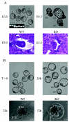
FIG. 2.
E4F disruption results in early embryonic lethality. (A) The appearance of mutant embryos. Upper panels, E3.5 and E4.5 embryos were flushed out from the uteri and were photographed under bright field conditions. E3.5 blastocysts were subsequently genotyped by PCR. Note normal appearance of mutant embryos at E3.5 and severe growth retardation at E4.5. Lower panels, the appearance of E5.5 embryos developing in utero, as revealed by hematoxylin and eosin staining of histologic sections. A typical picture of a WT and of a not yet fully resorbed E4F−/− embryo is shown. (B) Impaired in vitro development of E4F-deficient embryos. Blastocyst stage embryos were flushed from the uterus at E3.5, cultured in vitro for several days, and subsequently genotyped by PCR. While all the littermates displayed similar morphology at E3.5 (upper left panel), E4F−/− embryos appeared growth retarded after 24 h of culture (upper right panel). After 72 h of in vitro culture, WT and E4F+/− embryos developed outgrowths composed of the inner cell mass (ICM) surrounded by a single layer of trophoblast giant cells (TGC) (lower left panel), whereas E4F−/− embryos degenerated inside the zonae pellucidae (lower right panel).
FIG. 3.
FIG. 3.
S-phase progression in E4F-deficient embryos. (A) S-phase progression was gauged by determining BrdU incorporation in blastocysts recovered from E4F+/− intercrosses. Embryos were cultured in vitro for 24 h, pulsed for 20 min with BrdU, and processed for BrdU immunostaining and DAPI counterstaining. (B) Series of Z-plane images were stacked and analyzed by deconvolution microscopy to precisely quantify the percentage of BrdU-positive cells in WT and E4F−/− blastocysts after 24 h of culture. Error bars indicate standard deviations.
FIG. 4.
FIG. 4.
M-phase progression defects in E4F-deficient embryos. (A) Increased mitotic index in E4F−/− embryos. Shown are representative WT and mutant littermates that were cultured in vitro for 24 h and then fixed and stained with antibody against phospho-(Ser 10) histone H3 (HH3 P-Ser10), a marker of mitotic cells. Nuclei were counterstained with DAPI. (B) The mitotic index in freshly isolated E3.5 embryos (T = 0) or in embryos cultured in vitro for 24 h (T = 24) was calculated as the mean number of HH3 P-Ser10-positive cells per embryo divided by the mean number of cells per embryo, times 100% (n = 15 for each genotype). (C and D) E4F−/− cells are blocked at the prometaphase stage. (C) Typical prometaphase figures (arrows) observed after DAPI staining of cultured E4F-deficient embryos. (D) Mitotic figures were identified in cultured WT and E4F−/− embryos based on the HH3 P-Ser10 immunostaining and were classified into the appropriate mitotic stage. The data are presented as percentages of these various stages among all M-phase cells. (E) Defective mitotic exit in E4F−/− embryos. Embryos were isolated at E3.5, cultured for 24 h, gamma irradiated, and left in culture for six additional hours. Embryos were then fixed, processed for the HH3 P-Ser10 immunostaining, and subsequently lysed individually for genotyping by PCR analysis. The number of mitotic cells (HH3 P-Ser10 positive) in nonirradiated (black circles) or irradiated embryos (open circles) is plotted according to their genotype.
FIG. 5.
FIG. 5.
Embryonic cells that lack E4F are arrested in prometaphase with an active spindle checkpoint, as revealed by Bub1 and BubR1 immunostaining. Examples of WT embryos, representing various phases of the M-phase progression, and representative E4F-deficient embryos (KO) were costained in panel A with anti-HH3 P-Ser10 antibodies, anti-Bub1 antibodies, and DAPI, or in panel B, with anti-HH3 P-Ser10 antibodies, anti-BubR1 antibodies, and DAPI.
FIG. 6.
FIG. 6.
E4F is localized on the mitotic spindle. WT blastocysts were immunostained with a specific anti-E4F antibody. The mitotic spindle was visualized by α-tubulin immunostaining. Nuclei were counterstained with DAPI.
FIG. 7.
FIG. 7.
E4F−/− embryos exhibit abnormal mitotic figures and increased cell death. (A) Mitotic cells in in vitro-cultured blastocysts were identified by HH3 P-Ser10 immunostaining. Examples of abnormal mitotic figures seen in E4F-deficient embryos are shown. Arrows indicate misaligned chromosomes. (B) Increased apoptosis in E4F-deficient embryos. Merged image obtained from representative WT and E4F−/− blastocysts cultured for 24 h and processed for DAPI and TUNEL staining.

References

    1. Cleveland, D. W., Y. Mao, and K. F. Sullivan. 2003. Centromeres and kinetochores: from epigenetics to mitotic checkpoint signaling. Cell 112:407-421. - PubMed
    1. Colombo, R., G. F. Draetta, and S. Chiocca. 2003. Modulation of p120E4F transcriptional activity by the Gam1 adenoviral early protein. Oncogene 22:2541-2547. - PubMed
    1. Cutts, S. M., K. J. Fowler, B. T. Kile, L. L. Hii, R. A. O'Dowd, D. F. Hudson, R. Saffery, P. Kalitsis, E. Earle, and K. H. Choo. 1999. Defective chromosome segregation, microtubule bundling and nuclear bridging in inner centromere protein gene (Incenp)-disrupted mice. Hum. Mol. Genet. 8:1145-1155. - PubMed
    1. Fajas, L., C. Paul, A. Vie, S. Estrach, R. Medema, J. M. Blanchard, C. Sardet, and M. L. Vignais. 2001. Cyclin A is a mediator of p120E4F-dependent cell cycle arrest in G1. Mol. Cell. Biol. 21:2956-2966. - PMC - PubMed
    1. Fajas, L., C. Paul, O. Zugasti, L. Le Cam, J. Polanowska, E. Fabbrizio, R. Medema, M. L. Vignais, and C. Sardet. 2000. pRB binds to and modulates the transrepressing activity of the E1A-regulated transcription factor p120E4F. Proc. Natl. Acad. Sci. USA 97:7738-7743. - PMC - PubMed

Publication types

MeSH terms