

Clinical implication of expression of vascular endothelial growth factor-C in metastatic lymph nodes of uterine cervical cancers
- PMID: 15226772
- PMCID: PMC2409847
- DOI: 10.1038/sj.bjc.6601963
Clinical implication of expression of vascular endothelial growth factor-C in metastatic lymph nodes of uterine cervical cancers
Abstract
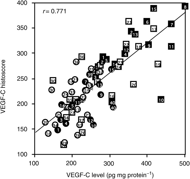
Vascular endothelial cell growth factor (VEGF)-C levels were significantly (P<0.05) increased in 24 out of 40 metastatic lymph node lesions of uterine cervical cancers. The prognosis of the 24 patients with increased VEGF-C level in metastatic lymph node lesions was poor and the 24-month survival rate was 38%, while the rate of the 16 patients with no change in VEGF-C level in metastatic lymph node lesions was 81%. There was a significant (P<0.01) difference in the 24-month survival rates between the two groups. This is novel, direct evidence that VEGF-C might contribute to the aggressive lymphangitic metastasis in uterine cervical cancers, and that the increase in VEGF-C level from primary tumour to metastatic lymph node might be a prognostic indicator.
Figures


Similar articles
-
Clinical implication of expression of platelet-derived endothelial cell growth factor (PD-ECGF) in metastatic lesions of uterine cervical cancers.Cancer Res. 1999 Jul 1;59(13):3041-4. Cancer Res. 1999. PMID: 10397240
-
Increased expression of vascular endothelial growth factor C in papillary thyroid carcinoma correlates with cervical lymph node metastases.Clin Cancer Res. 2005 Nov 15;11(22):8063-9. doi: 10.1158/1078-0432.CCR-05-0646. Clin Cancer Res. 2005. PMID: 16299237
-
CD44v3 and VEGF-C expression and its relationship with lymph node metastasis in squamous cell carcinomas of the uterine cervix.Asian Pac J Cancer Prev. 2014;15(12):5049-53. doi: 10.7314/apjcp.2014.15.12.5049. Asian Pac J Cancer Prev. 2014. PMID: 24998585
-
Elevated expression of thymosin β4, vascular endothelial growth factor (VEGF), and hypoxia inducible factor (HIF)-1α in early-stage cervical cancers.Pathol Oncol Res. 2011 Sep;17(3):493-502. doi: 10.1007/s12253-010-9327-x. Epub 2011 Jan 7. Pathol Oncol Res. 2011. PMID: 21213129
-
Vascular endothelial growth factor C expression is closely correlated with lymph node recurrence and poor prognosis in patients with early stage cervical cancer.J Int Med Res. 2013 Oct;41(5):1541-9. doi: 10.1177/0300060513493038. Epub 2013 Aug 20. J Int Med Res. 2013. PMID: 23963849
Cited by
-
Clinicopathological Significance of VEGF-C, VEGFR-3 and Cyclooxygenase-2 in Early-Stage Cervical Cancer.Int J Biomed Sci. 2008 Mar;4(1):58-63. Int J Biomed Sci. 2008. PMID: 23675067 Free PMC article.
-
VEGF-C/Flt-4 axis in tumor cells contributes to the progression of oral squamous cell carcinoma via upregulating VEGF-C itself and contactin-1 in an autocrine manner.Am J Cancer Res. 2018 Oct 1;8(10):2046-2063. eCollection 2018. Am J Cancer Res. 2018. PMID: 30416855 Free PMC article.
-
The potential applications of T cell receptor (TCR)-like antibody in cervical cancer immunotherapy.Hum Vaccin Immunother. 2021 Sep 2;17(9):2981-2994. doi: 10.1080/21645515.2021.1913960. Epub 2021 May 14. Hum Vaccin Immunother. 2021. PMID: 33989511 Free PMC article. Review.
-
Prognostic role of vascular endothelial growth factor in cervical cancer: a meta-analysis.Oncotarget. 2017 Apr 11;8(15):24797-24803. doi: 10.18632/oncotarget.15044. Oncotarget. 2017. PMID: 28177889 Free PMC article.
-
Resistance Mechanisms to Anti-angiogenic Therapies in Cancer.Front Oncol. 2020 Feb 27;10:221. doi: 10.3389/fonc.2020.00221. eCollection 2020. Front Oncol. 2020. PMID: 32175278 Free PMC article. Review.
References
-
- Amioka T, Kitadai Y, Tanaka S, Haruma K, Yoshihara M, Yasui W, Chayama K (2002) Vascular endothelial growth factor-C expression predicts lymph node metastasis of human gastric carcinomas invading the submucosa. Eur J Cancer 38: 1413–1419 - PubMed
-
- Beasley NJ, Prevo R, Banerji S, Leek RD, Moore J, van Trappen P, Cox G, Harris AL, Jackson DG (2002) Intratumoral lymphangiogenesis and lymph node metastasis in head and neck cancer. Cancer Res 62: 1315–1320 - PubMed
-
- Bradford M (1976) A rapid and sensitive method for the quantitation of microgram quantities of protein utilizing the principle of protein-dye binding. Anal Biochem 72: 315–323 - PubMed
-
- Chen RJ, Chang DY, Yen ML, Lee EF, Huang SC, Chow SN Hsieh CY (1998) Prognostic factors of primary adenocarcinoma of the uterine cervix. Gynecol Oncol 69: 157–164 - PubMed
-
- FIGO News (1989) Int J Gynecol Obstet 28: 189–193
