

Hippocampal synapses depend on hippocampal estrogen synthesis
- PMID: 15229239
- PMCID: PMC6729232
- DOI: 10.1523/JNEUROSCI.5186-03.2004
Hippocampal synapses depend on hippocampal estrogen synthesis
Abstract
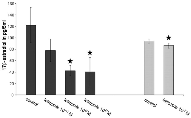
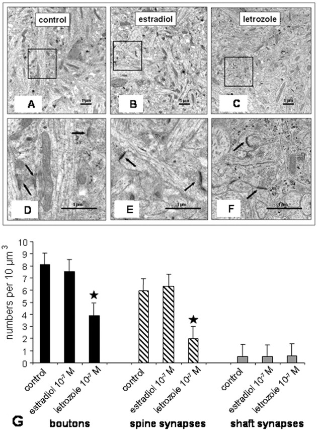
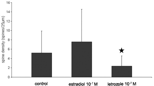
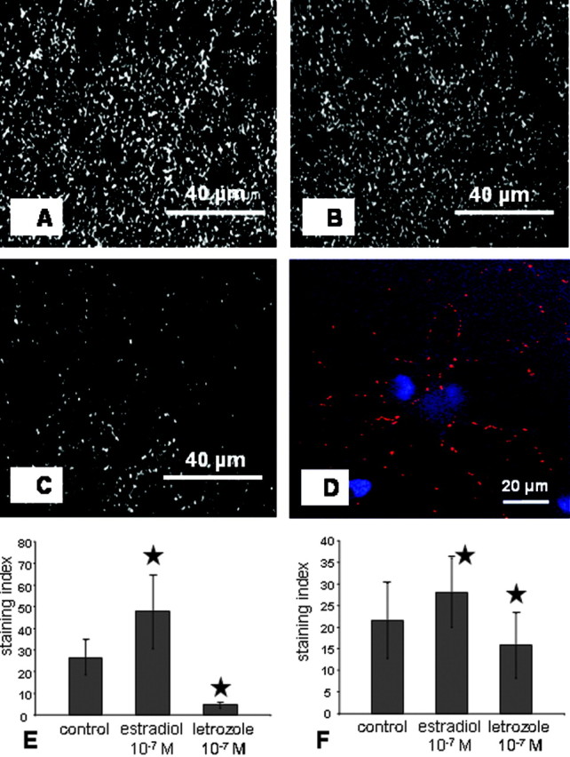
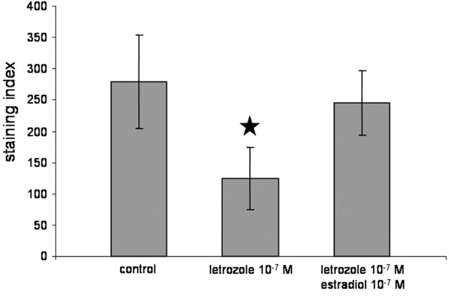
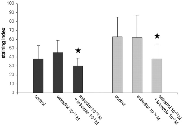
Estrogens have been described to induce synaptogenesis in principal neurons of the hippocampus and have been shown to be synthesized and released by exactly these neurons. Here, we have focused on the significance of local estrogen synthesis on spine synapse formation and the synthesis of synaptic proteins. To this end, we reduced hippocampal estrogen synthesis in vitro with letrozole, a reversible nonsteroidal aromatase inhibitor. In hippocampal slice cultures, letrozole treatment resulted in a dose-dependent decrease of 17beta-estradiol as quantified by RIA. This was accompanied by a significant decrease in the density of spine synapses and in the number of presynaptic boutons. Quantitative immunohistochemistry revealed a downregulation of spinophilin, a marker of dendritic spines, and synaptophysin, a protein of presynaptic vesicles, in response to letrozole. Surprisingly, no increase in the density of spines, boutons, and synapses and in spinophilin expression was seen after application of estradiol to the medium of cultures that had not been treated with letrozole. However, synaptophysin expression was upregulated under these conditions. Our results point to an essential role of endogenous hippocampal estrogen synthesis in the maintenance of hippocampal spine synapses.
Figures

Similar articles
-
Neurosteroid synthesis in the hippocampus: role in synaptic plasticity.Neuroscience. 2005;136(3):833-42. doi: 10.1016/j.neuroscience.2005.03.056. Neuroscience. 2005. PMID: 16344155
-
Inhibition of hippocampal estrogen synthesis causes region-specific downregulation of synaptic protein expression in hippocampal neurons.Hippocampus. 2006;16(5):464-71. doi: 10.1002/hipo.20173. Hippocampus. 2006. PMID: 16502389
-
Direct and indirect effects of estrogen on rat hippocampus.Neuroscience. 2006;138(3):765-72. doi: 10.1016/j.neuroscience.2005.05.061. Epub 2005 Dec 1. Neuroscience. 2006. PMID: 16324798 Review.
-
Cholesterol-promoted synaptogenesis requires the conversion of cholesterol to estradiol in the hippocampus.Hippocampus. 2009 Aug;19(8):692-705. doi: 10.1002/hipo.20548. Hippocampus. 2009. PMID: 19156851
-
Estrogen-regulated synaptogenesis in the hippocampus: sexual dimorphism in vivo but not in vitro.J Steroid Biochem Mol Biol. 2012 Aug;131(1-2):24-9. doi: 10.1016/j.jsbmb.2011.11.010. Epub 2011 Nov 25. J Steroid Biochem Mol Biol. 2012. PMID: 22138012 Review.
Cited by
-
Sex as a Determinant of Age-Related Changes in the Brain.Int J Mol Sci. 2024 Jun 28;25(13):7122. doi: 10.3390/ijms25137122. Int J Mol Sci. 2024. PMID: 39000227 Free PMC article. Review.
-
Agomelatine (S20098) modulates the expression of cytoskeletal microtubular proteins, synaptic markers and BDNF in the rat hippocampus, amygdala and PFC.Psychopharmacology (Berl). 2012 Jun;221(3):493-509. doi: 10.1007/s00213-011-2597-5. Epub 2011 Dec 8. Psychopharmacology (Berl). 2012. PMID: 22160164
-
Estradiol meets notch signaling in developing neurons.Front Endocrinol (Lausanne). 2011 Aug 11;2:21. doi: 10.3389/fendo.2011.00021. eCollection 2011. Front Endocrinol (Lausanne). 2011. PMID: 22654797 Free PMC article.
-
De Novo Synthesized Estradiol: A Role in Modulating the Cerebellar Function.Int J Mol Sci. 2020 May 7;21(9):3316. doi: 10.3390/ijms21093316. Int J Mol Sci. 2020. PMID: 32392845 Free PMC article. Review.
-
Aromatase promoter I.f is regulated by estrogen receptor alpha (ESR1) in mouse hypothalamic neuronal cell lines.Biol Reprod. 2009 Nov;81(5):956-65. doi: 10.1095/biolreprod.109.077206. Epub 2009 Jul 15. Biol Reprod. 2009. PMID: 19605792 Free PMC article.
References
-
- Abdelgadir ES, Resko JA, Ojeda SR, Lephart ED, McPhaul MJ, Roselli CE (1994) Androgens regulate aromatase cytochrome P450 messenger ribonucleic acid in rat brain. Endocrinology 135: 395-401. - PubMed
-
- Azcoitia I, Sierra A, Garcia-Segura LM (1999) Localization of estrogen receptor β-immunoreactivity in astrocytes of the adult rat brain. Glia 26: 260-267. - PubMed
-
- Azcoitia I, Sierra A, Veiga S, Honda S, Harada N, Garcia-Segura LM (2001) Brain aromatase is neuroprotective. J Neurobiol 47: 318-329. - PubMed
Publication types
MeSH terms
Substances
LinkOut - more resources
Full Text Sources