. 2004 Jun 30;24(26):6003-10.
doi: 10.1523/JNEUROSCI.4848-03.2004.

Spike synchronization in the cortex/basal-ganglia networks of Parkinsonian primates reflects global dynamics of the local field potentials

Affiliations

Spike synchronization in the cortex/basal-ganglia networks of Parkinsonian primates reflects global dynamics of the local field potentials

Joshua A Goldberg et al. J Neurosci. .

Abstract

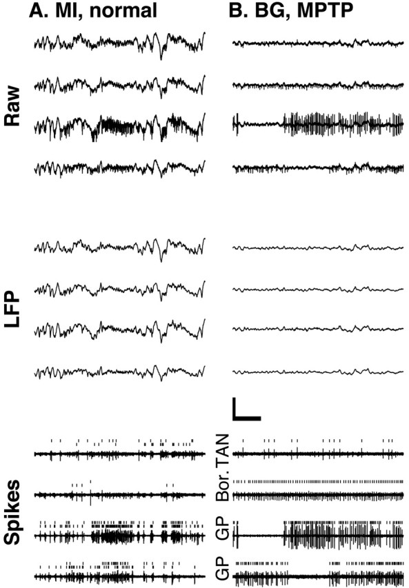
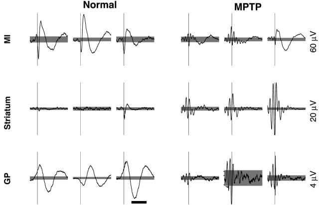
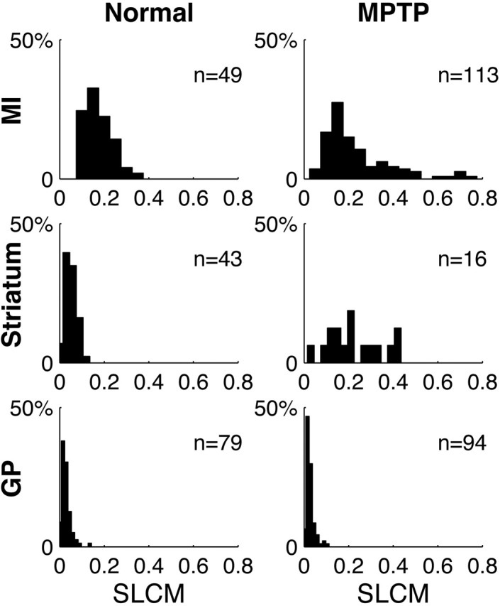
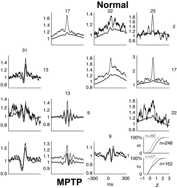
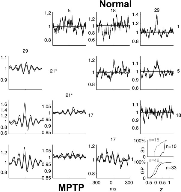
Cortical local field potentials (LFPs) reflect synaptic potentials and accordingly correlate with neuronal discharge. Because LFPs are coherent across substantial cortical areas, we hypothesized that cortical spike correlations could be predicted from them. Because LFPs recorded in the basal ganglia (BG) are also correlated with neuronal discharge and are clinically accessible in Parkinson's disease patients, we were interested in testing this hypothesis in the BG, as well. We recorded LFPs and unit discharge from multiple electrodes, which were placed in primary motor cortex or in the basal ganglia (striatum and pallidum) of two monkeys before and after rendering them parkinsonian with 1-methyl-4-phenyl-1,2,3,6-tetrahydropyridine. We used the method of partial spectra to construct LFP predictors of the spike cross-correlation functions (CCFs). The predicted CCF is an estimate of the correlation between two neurons under the assumption that their association is explained solely by the association of each with the LFP recorded on a third electrode. In the normal condition, the predictors account for cortical rate covariations but not for the association among the tonically active neurons of the striatum. In the parkinsonian condition, with the appearance of 10 Hz oscillations throughout the cortex-basal ganglia networks, the LFP predictors account remarkably better for the CCFs in both the cortex and the basal ganglia. We propose that, in the parkinsonian condition, the cortex-basal ganglia networks are more tightly related to global modes of brain dynamics that are echoed in the LFP.


Figures

Figure 1.
Figure 1.
Multiple-electrode recordings from the MI and the BG. Four simultaneous 3 sec electrode traces of the extracellular recording from monkey S are shown. Raw, The output of the amplifiers, which includes both spiking activity and the slower LFPs. LFP, Digitally low-pass filtered (<150 Hz) versions of the raw traces. Calibration: 500 msec, 500 μV. Spikes, Digitally high-pass filtered versions (>500 Hz) of the corresponding raw data (arbitrary y scale). Ticks mark the spikes identified by the on-line spike sorting. Two units are shown in each MI trace. Traces in A are from MI in the normal condition. Traces in B are from the BG in the MPTP-treated condition. GP, Neuron recorded from the globus pallidus. Bor., Border cell located on the border between the striatum and the GP. TAN, A tonically active neuron of the striatum.
Figure 2.
Figure 2.
STAs of the LFP. Three examples of the STAs from each structure in both the normal and the MPTP-treated condition. Calibration: 500 msec. Thin vertical lines mark the time of the spikes. The scales of the vertical lines are given to the right of the examples. Gray background, Confidence interval of 99.9%. Examples from the primary motor cortex in the MPTP condition are taken from monkey Z. All other examples are from monkey S. In the rightmost columns in both conditions (Normal, MPTP), the spike and LFP are recorded on the same electrode.
Figure 3.
Figure 3.
Distribution of the SLCMs between spikes and the LFP. Frequency histograms of the SLCMs from all structures in both the normal and the MPTP-treated conditions in monkey S. The number (n) of units used to generate the histogram is indicated.
Figure 4.
Figure 4.
Comparison of predicted and observed CCFs in MI in the normal and MPTP-treated conditions. Top triangular matrix, Observed (black) and predicted (gray) CCFs of all pairs from four simultaneously recorded units in the normal condition. Bottom triangular matrix, Observed and predicted CCFs in the MPTP-treated condition. CCFs are normalized so that they tend to unity for large absolute time lags. Inset, Cumulative frequency distributions of the Fisher Z-transformed value (z) of Pearson's product-moment correlation-coefficient between the predicted and observed CCFs in the normal (gray) and MPTP-treated (black) conditions, calculated for the cases in which the observed CCFs was significantly modulated. S and Z denote the monkey. The number of neuronal pairs (n) used to generate the histogram is indicated. The significant (p < 0.05; two-tailed Wilcoxon rank-sum test) shift in the histograms indicates that the value of z is stochastically larger in the MPTP condition, implying a closer match between the predicted and observed CCF.
Figure 5.
Figure 5.
Comparison of predicted and observed CCFs in the BG in the normal and MPTP-treated conditions. Format is the same as in Figure 4. All units are striatal TANs, except unit 21 (marked by an asterisk) in the MPTP condition, which is a GP unit. Inset, The cumulative frequency histograms are calculated separately for striatal (Str.) and GP pairs. The value of z is also stochastically larger when all BG pairs are pooled together (p < 0.001; two-tailed Wilcoxon rank-sum test), implying a closer match between the predicted and observed CCF in the MPTP condition.

References

    1. Abeles M (1974) A journey into the brain. In: Signal analysis and pattern recognition in biomedical engineering (Inbar GF, ed), pp 41-59. New York: Wiley.
    1. Abeles M (1982) Local cortical circuits. (Braitenberg V, Barlow HB, Bullock H, Florey E, Grüsser O-J, Peters A, eds). New York: Springer.
    1. Aosaki T, Kimura M, Graybiel AM (1995) Temporal and spatial characteristics of tonically active neurons of the primate's striatum. J Neurophysiol 73: 1234-1252. - PubMed
    1. Arieli A, Sterkin A, Grinvald A, Aertsen A (1996) Dynamics of ongoing activity: explanation of the large variability in evoked cortical responses. Science 273: 1868-1871. - PubMed
    1. Asanuma H (1989) The motor cortex. New York: Raven.

Publication types

LinkOut - more resources