

Bmx tyrosine kinase transgene induces skin hyperplasia, inflammatory angiogenesis, and accelerated wound healing
- PMID: 15229285
- PMCID: PMC515354
- DOI: 10.1091/mbc.e04-03-0241
Bmx tyrosine kinase transgene induces skin hyperplasia, inflammatory angiogenesis, and accelerated wound healing
Abstract
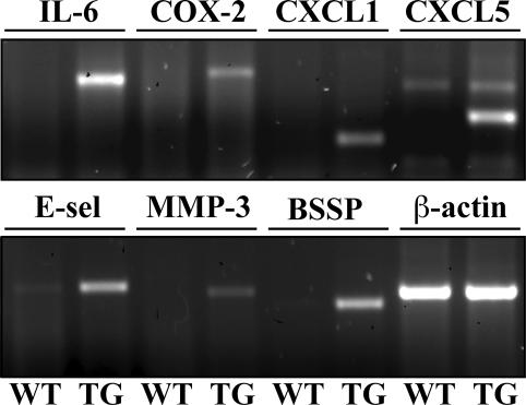
The Bmx gene, a member of the Tec family of nonreceptor protein tyrosine kinases, is expressed in arterial endothelium and in certain hematopoietic and epithelial cells. Previous in vitro studies have implicated Bmx signaling in cell migration and survival and suggested that it contributes to the progression of prostate carcinomas. However, the function of Bmx in normal tissues in vivo is unknown. We show here that Bmx expression is induced in skin keratinocytes during wound healing. To analyze the role of Bmx in epidermal keratinocytes in vivo, we generated transgenic mice overexpressing Bmx in the skin. We show that Bmx overexpression accelerates keratinocyte proliferation and wound reepithelialization. Bmx expression also induces chronic inflammation and angiogenesis in the skin, and gene expression profiling suggests that this occurs via cytokine-mediated recruitment of inflammatory cells. Our studies provide the first data on Bmx function in vivo and form the basis of evaluation of its role in epithelial neoplasia.
Figures

Similar articles
-
Deletion of the endothelial Bmx tyrosine kinase decreases tumor angiogenesis and growth.Cancer Res. 2012 Jul 15;72(14):3512-21. doi: 10.1158/0008-5472.CAN-11-1070. Epub 2012 May 16. Cancer Res. 2012. PMID: 22593188
-
VEGF/VPF overexpression in skin of transgenic mice induces angiogenesis, vascular hyperpermeability and accelerated tumor development.Oncogene. 1998 Jul 23;17(3):303-11. doi: 10.1038/sj.onc.1201928. Oncogene. 1998. PMID: 9690512
-
Bmx tyrosine kinase has a redundant function downstream of angiopoietin and vascular endothelial growth factor receptors in arterial endothelium.Mol Cell Biol. 2001 Jul;21(14):4647-55. doi: 10.1128/MCB.21.14.4647-4655.2001. Mol Cell Biol. 2001. PMID: 11416142 Free PMC article.
-
BMX and its role in inflammation, cardiovascular disease, and cancer.Int Rev Immunol. 2012 Apr;31(2):166-73. doi: 10.3109/08830185.2012.663838. Int Rev Immunol. 2012. PMID: 22449076 Review.
-
The wound repair-associated keratins 6, 16, and 17. Insights into the role of intermediate filaments in specifying keratinocyte cytoarchitecture.Subcell Biochem. 1998;31:173-204. Subcell Biochem. 1998. PMID: 9932493 Review. No abstract available.
Cited by
-
Nuclear localization of the tyrosine kinase BMX mediates VEGFR2 expression.J Cell Mol Med. 2020 Jan;24(1):126-138. doi: 10.1111/jcmm.14663. Epub 2019 Oct 23. J Cell Mol Med. 2020. PMID: 31642192 Free PMC article.
-
Transcriptome analysis during photostimulated recrudescence reveals distinct patterns of gene regulation in Siberian hamster ovaries†.Biol Reprod. 2020 Mar 13;102(3):539-559. doi: 10.1093/biolre/ioz210. Biol Reprod. 2020. PMID: 31724051 Free PMC article.
-
Tyrosine kinase BMX phosphorylates phosphotyrosine-primed motif mediating the activation of multiple receptor tyrosine kinases.Sci Signal. 2013 May 28;6(277):ra40. doi: 10.1126/scisignal.2003936. Sci Signal. 2013. PMID: 23716717 Free PMC article.
-
Compensatory upregulation of tyrosine kinase Etk/BMX in response to androgen deprivation promotes castration-resistant growth of prostate cancer cells.Cancer Res. 2010 Jul 1;70(13):5587-96. doi: 10.1158/0008-5472.CAN-09-4610. Epub 2010 Jun 22. Cancer Res. 2010. PMID: 20570899 Free PMC article.
-
Location and level of Etk expression in neurons are associated with varied severity of traumatic brain injury.PLoS One. 2012;7(6):e39226. doi: 10.1371/journal.pone.0039226. Epub 2012 Jun 18. PLoS One. 2012. PMID: 22723969 Free PMC article.
References
-
- Abassi, Y.A., Rehn, M., Ekman, N., Alitalo, K., and Vuori, K. (2003). p130Cas couples the tyrosine kinase Bmx/Etk with regulation of the actin cytoskeleton and cell migration. J. Biol. Chem. 278, 35636–35643. - PubMed
-
- Bagheri-Yarmand, R., Mandal, M., Taludker, A.H., Wang, R.A., Vadlamudi, R.K., Kung, H.J., and Kumar, R. (2001). Etk/Bmx tyrosine kinase activates Pak1 and regulates tumorigenicity of breast cancer cells. J. Biol. Chem. 276, 29403–29409. - PubMed
-
- Belperio, J.A., Keane, M.P., Arenberg, D.A., Addison, C.L., Ehlert, J.E., Burdick, M.D., and Strieter, R.M. (2000). CXC chemokines in angiogenesis. J. Leukoc. Biol. 68, 1–8. - PubMed
-
- Breitenbach, U., Tuckermann, J.P., Bebhardt, C., Richter, K.H., Furstenberger, G., Christofori, G., and Angel, P. (2001). Keratinocyte-specific onset of serine protease BSSP expression in experimental carcinogenesis. J. Invest. Dermatol. 117, 634–640. - PubMed
-
- Chau, C.H., Chen, K.Y., Deng, H.T., Kim, K.J., Hosoya, K., Terasaki, T., Shih, H.M., and Ann, D.K. (2002). Coordinating Etk/Bmx activation and VEGF upregulation to promote cell survival and proliferation. Oncogene 21, 8817–8829. - PubMed
Publication types
MeSH terms
Substances
LinkOut - more resources
Full Text Sources
Molecular Biology Databases
Miscellaneous
