

Case Reports
doi: 10.1056/NEJMoa032655.
Fatal myositis due to the microsporidian Brachiola algerae, a mosquito pathogen
Affiliations
- PMID: 15229306
- PMCID: PMC3109631
- DOI: 10.1056/NEJMoa032655
Item in Clipboard
Case Reports
Fatal myositis due to the microsporidian Brachiola algerae, a mosquito pathogen
N Engl J Med.
.
No abstract available
Figures
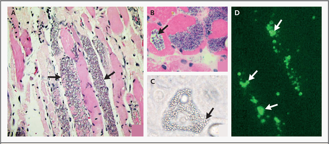
In Panel A, a low-power photomicrograph of the initial muscle-biopsy specimen obtained after the patient presented with myositis and muscle pain shows multiple organisms in the muscle fibers (arrows) with associated cell lysis but little or no inflammation (hematoxylin and eosin, ×100). In Panel B, a high-power photomicrograph of the second muscle-biopsy specimen demonstrates cytolysis (arrow) of the muscle fibers surrounding spores (×400). In Panel C, a phase-contrast photomicrograph of B. algerae isolated from muscle shows spores (arrow) and proliferative forms throughout the cytoplasm of the host cell (×400). In Panel D, a high-power photomicrograph shows clear immunofluorescence staining (arrows) of the initial muscle-biopsy specimen with antiserum against B. algerae (human corneal isolate) (×400).
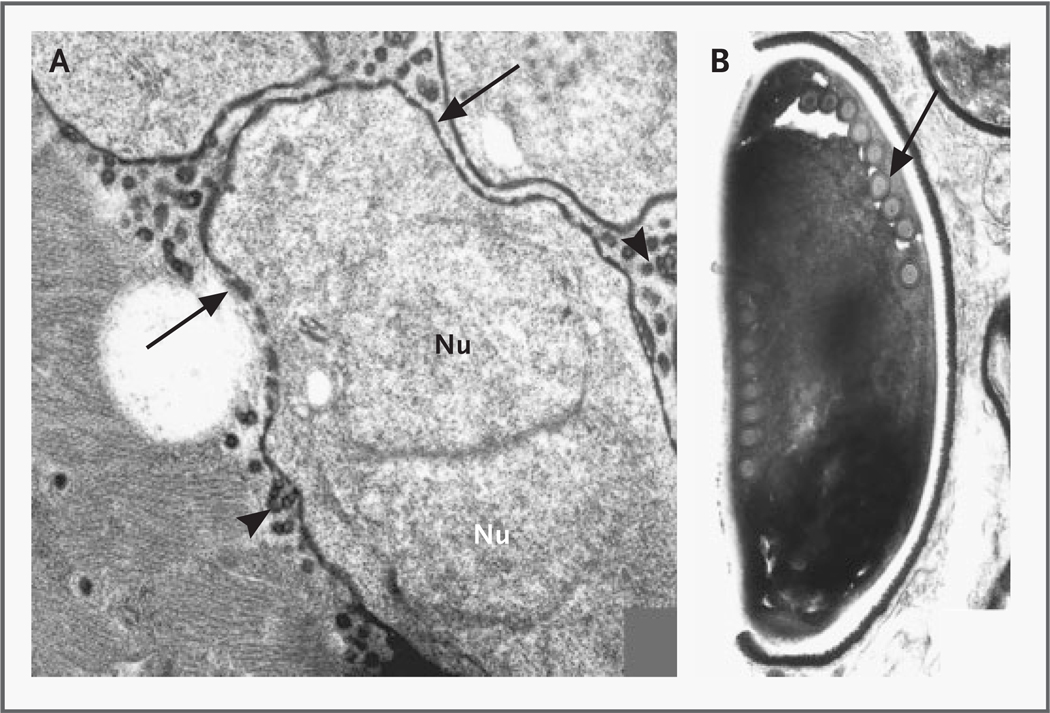
In Panel A, a diplokaryon, a thickened plasmalemma (arrows), and vesicotubular extensions (arrowheads) are evident in proliferative forms of Brachiola algerae in muscle (×14,000). Nu denotes nucleus. Panel B shows a mature B. algerae spore with a single row of nine polar filaments (arrow) in cross section. Mature spores in this biopsy specimen had only single rows of 8 to 12 polar filaments in cross section (×18,300).
Similar articles
-
Anncaliia algerae microsporidial myositis.Emerg Infect Dis. 2014 Feb;20(2):185-91. doi: 10.3201/eid2002.131126. Emerg Infect Dis. 2014. PMID: 24447398 Free PMC article.
-
Brachiola vesicularum, n. g., n. sp., a new microsporidium associated with AIDS and myositis.J Eukaryot Microbiol. 1998 May-Jun;45(3):240-51. doi: 10.1111/j.1550-7408.1998.tb04532.x. J Eukaryot Microbiol. 1998. PMID: 9627985
-
An analysis of the microsporidian genus Brachiola, with comparisons of human and insect isolates of Brachiola algerae.J Eukaryot Microbiol. 2004 Nov-Dec;51(6):678-85. doi: 10.1111/j.1550-7408.2004.tb00608.x. J Eukaryot Microbiol. 2004. PMID: 15666726 Free PMC article.
-
Treatment of microsporidiosis including albendazole.Parasitol Res. 2003 Jun;90 Supp 1:S14-8. doi: 10.1007/s00436-002-0753-x. Epub 2002 Dec 10. Parasitol Res. 2003. PMID: 12811545 Review.
-
Microsporidial disease in HIV-infected patients: a report of 42 patients and review of the literature.Scand J Infect Dis. 1998;30(4):331-8. doi: 10.1080/00365549850160594. Scand J Infect Dis. 1998. PMID: 9817510 Review.
Cited by
-
MicroAnnot: A Dedicated Workflow for Accurate Microsporidian Genome Annotation.Int J Mol Sci. 2024 Jan 10;25(2):880. doi: 10.3390/ijms25020880. Int J Mol Sci. 2024. PMID: 38255958 Free PMC article.
-
Infective myositis.Brain Pathol. 2021 May;31(3):e12950. doi: 10.1111/bpa.12950. Brain Pathol. 2021. PMID: 34043257 Free PMC article. Review.
-
Microsporidia-Emergent Pathogens in the Global Food Chain (Trends in Parasitology 32, 336-348; April 2, 2016).Trends Parasitol. 2016 Aug;32(8):657. doi: 10.1016/j.pt.2016.06.002. Epub 2016 Jun 27. Trends Parasitol. 2016. PMID: 27365191 No abstract available.
-
An Ultrastructural Study of the Extruded Polar Tube of Anncaliia algerae (Microsporidia).J Eukaryot Microbiol. 2020 Jan;67(1):28-44. doi: 10.1111/jeu.12751. Epub 2019 Aug 8. J Eukaryot Microbiol. 2020. PMID: 31332877 Free PMC article.
-
Small-molecule screen in C. elegans identifies benzenesulfonamides as inhibitors of microsporidia spores.NPJ Antimicrob Resist. 2025 May 21;3(1):41. doi: 10.1038/s44259-025-00116-0. NPJ Antimicrob Resist. 2025. PMID: 40399462 Free PMC article.
References
-
- Wittner M, Weiss LM, editors. The microsporidia and microsporidiosis. Washington, D.C.: ASM Press; 1999.
-
- Lipsky PE, van der Heijde DMFM, St Clair EW, et al. Infliximab and methotrexate in the treatment of rheumatoid arthritis. N Engl J Med. 2000;343:1594–1602. - PubMed
-
- Visvesvara GS, Belloso M, Moura H, et al. Isolation of Nosema algerae from the cornea of an immunocompetent patient. J Eukaryot Microbiol. 1999;46:10S. - PubMed
-
- Weiss LM. Molecular phylogeny and diagnostic approaches to microsporidia. Contrib Microbiol. 2000;6:209–235. - PubMed
-
- Lowman PM, Takvorian PM, Cali A. The effects of elevated temperatures and various time-temperature combinations on the development of Brachiola (Nosema) algerae N. Comb. in mammalian cell culture. J Eukaryot Microbiol. 2000;47:221–234. - PubMed
Publication types
MeSH terms
Substances
Grants and funding
LinkOut - more resources
Full Text Sources
Medical
Molecular Biology Databases