. 2004 Jul 1:4:24.
doi: 10.1186/1471-2180-4-24.

Chlamydial development is blocked in host cells transfected with Chlamydophila caviae incA

Affiliations

Chlamydial development is blocked in host cells transfected with Chlamydophila caviae incA

Damir Alzhanov et al. BMC Microbiol. .

Abstract

Background: Chlamydiae produce a set of proteins, termed Inc proteins, that are localized to the inclusion membrane and exposed to the host cell cytosol. Little information exists regarding the interaction of Inc proteins with the eukaryotic cell. To examine these interactions, Vaccinia virus vectors and mammalian plasmid-based systems were used to express inc genes in mammalian cells.

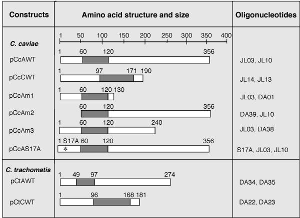
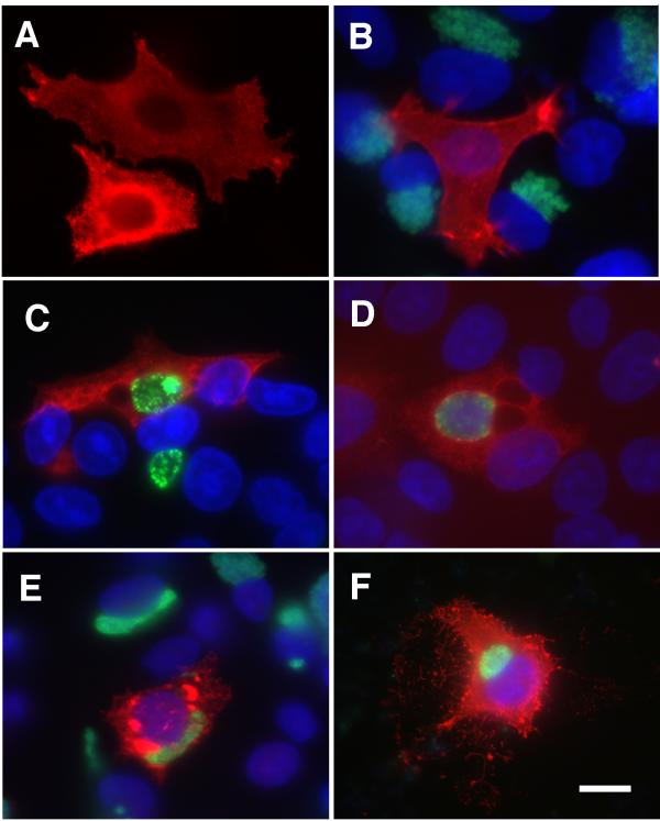
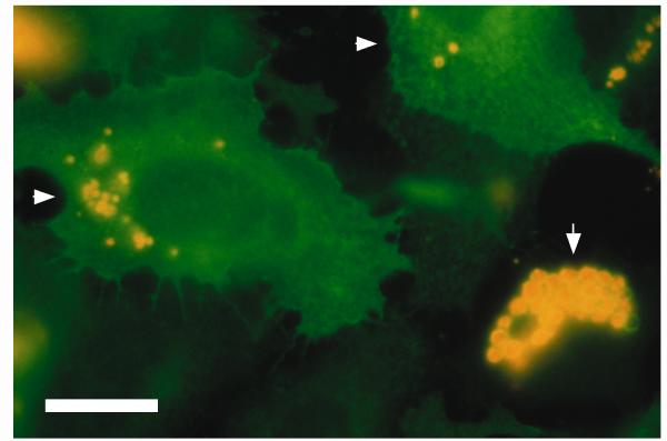
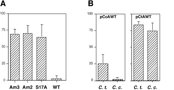
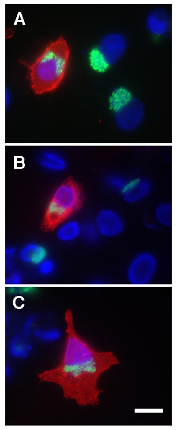
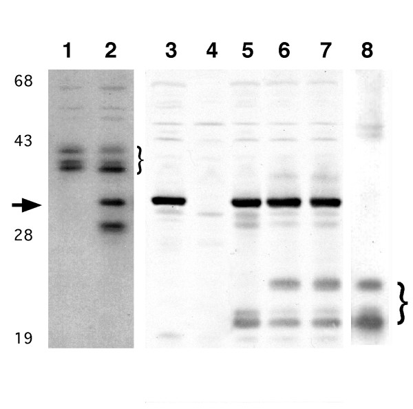
Results: Cells transfected with plasmids expressing Chlamydophila caviae incA were not productively infected by C. caviae. Expression of C. caviae incA also reduced inclusion formation by Chlamydia trachomatis, but not to the degree seen for C. caviae. Chlamydia trachomatis incA did not block development of either C. trachomatis or C. caviae. Deletion mutagenesis was used to demonstrate that plasmids encoding either the amino or carboxy-terminal regions of the protein, as well as the changing of a single amino acid within IncA (serine 17) could not block C. caviae infection. Immunoblot analysis of truncated IncA in a Vaccinia virus system provided evidence that serine 17 of C. caviae IncA is a target for phosphorylation.

Conclusions: These experiments provide insight into the interaction of Inc proteins with the host cell and introduce a model system where these interactions can be explored further.


Figures

Figure 1
Figure 1
Schematic representations of inc gene constructs used in this study. The plasmid name (left) and a schematic of the predicted protein structure (center), and oligonucleotides used in construction (right; Table 1) are indicated for each plasmid. The scale bar at top indicates the length in amino acids of each predicted gene product. All pcDNA4/HisMax C constructs encode a polyhistidine tag at the amino terminus. Chlamydophila caviae and C. trachomatis incA were also expressed via pcDNA3.1(+), in constructs that do not encode proteins with a polyhistidine tag. A large region of predicted hydrophobicity within each predicted protein is indicated for each construct (darker bar).
Figure 2
Figure 2
Effect of cytosolic expression of incA and incC from C. caviae or C. trachomatis. The vector pcDNA4/HisMaxC was used in each construct examined in this figure. IncA was detected by using specific monoclonal antibodies and labeled in red in each panel. Distribution of His/IncC fusion protein in the cytoplasm was detected indirectly by using anti polyhistidine tag monoclonal antibodies (Clontech) and also labeled in red. In panels B-F, the nuclei are labeled with DAPI (blue) and chlamydiae are labeled with anti-HSP60 (green). Panel A; HeLa cell transfected with pCcAWT (C. caviae incA). Panel B; HeLa cell transfected with pCcAWT in a monolayer subsequently infected at an MOI of 1.0 with C. caviae. Panel C; HeLa cell transfected with pCcAWT and subsequently infected at an MOI of 0.25 with C. trachomatis. Panel D; HeLa cell transfected with pCtAWT and subsequently infected at an MOI of 0.25 with C. trachomatis. Panel E; HeLa cell transfected with pCtAWT and subsequently infected at an MOI of 0.25 with C. caviae. Panel F; HeLa cell transfected with pCcCWT (C. caviae incC) and infected with C. caviae at MOI 1.0. The scale bar in F indicates 8 microns for each panel.
Figure 3
Figure 3
Examples of aberrant C. caviae inclusions formed in cells transfected with pcDN3.1(+) encoding C. caviae IncA. IncA is labeled green and chlamydial HSP60 is labeled yellow/orange in this image. Aberrant inclusions within transfected HeLa cells are shown with white arrowheads. A typical C. caviae inclusion within a nontransfected cell is shown in the low right corner of the figure (arrow). Cells were infected at a MOI of 0.5 and fixed with methanol 30 h post infection. The scale bar indicates 10 microns.
Figure 4
Figure 4
Quantitative analysis of transfection/infection experiments. The data shown represents the number of infected and transfected cells per one hundred transfected cells in the population. In all cases, cells to be used for quantification were infected at an MOI of 1 and were fixed for microscopy at 24 h post-infection. The error bars indicate standard deviations. Panel A; The relative infection efficiency of cells transfected with plasmids carrying each of the truncated or S17A mutated genes of C. caviae shown in Fig. 1 and Fig. 5, as compared to transfection with a plasmid expressing wild type (WT) of C. caviae incA. Panel B; Percent of cells infected with C. trachomatis (shown as C.t.) and C. caviae (shown as C.c.) after transfection with plasmids expressing either C. caviae or C. trachomatis incA (pCcAWT and pCtAWT, respectively).
Figure 5
Figure 5
Effect of cytosolic expression of deleted or mutated C. caviae incA sequences. Cells were transfected with either pCcAS17A, which encodes a protein differing from wild type at only a single amino acid (panel A), pCcAm2, which encodes a protein lacking the amino terminal 60 amino acids of IncA, (panel B), or pCcAM3, which encodes a protein lacking the carboxy terminal 116 amino acids of IncA (panel C). The fluorescence images are labeled with anti-polyhistidine monoclonal antibody (red), and anti-chlamydial HSP60 (green). The nuclei are labeled with DAPI (blue). The scale bar in C indicates 8 microns for each panel.
Figure 6
Figure 6
Immunoblots of lysates of HeLa cells infected with different Vaccinia virus (VV) recombinants. Full length (lanes 1 and 2) and truncated (lanes 5–8) IncA bands are indicated with brackets. Control VV-infected cells are in lane 3 and uninfected cells are in lane 4. As previously reported [4,8], full length C. caviae IncA migrates as three isoforms when produced during chlamydial infection (lane 1) or when expressed via a VV vector (lane 2). Lane 8 shows that truncated wild type IncA (residues 1 to 153) encoded by VV also is represented as three similar isoforms. Mutation of serines or threonines in the truncated incA are represented in lanes 5 (S17A), lane 6 (S121A) and lane 7 (T145A). Molecular mass standards are indicated in kilodaltons. The arrow indicates a background VV band recognized by the anti-incA antisera in some samples.

References

    1. Hackstadt T, Fischer ER, Scidmore MA, Rockey DD, Heinzen RA. Origins and functions of the chlamydial inclusion. Trends Microbiol. 1997;5:288–293. doi: 10.1016/S0966-842X(97)01061-5. - DOI - PubMed
    1. Bannantine JP, Rockey DD, Hackstadt T. Tandem genes of Chlamydia psittaci that encode proteins localized to the inclusion membrane. Molecular Microbiology. 1998;28:1017–1026. doi: 10.1046/j.1365-2958.1998.00867.x. - DOI - PubMed
    1. Bannantine JP, Stamm WE, Suchland RJ, Rockey DD. Chlamydia trachomatis IncA is localized to the inclusion membrane and is recognized by antisera from infected humans and primates. Infection and Immunity. 1998;66:6017–6021. - PMC - PubMed
    1. Rockey DD, Grosenbach D, Hruby DE, Peacock MG, Heinzen RA, Hackstadt T. Chlamydia psittaci IncA is phosphorylated by the host cell and is exposed on the cytoplasmic face of the developing inclusion. Mol Microbiol. 1997;24:217–228. doi: 10.1046/j.1365-2958.1997.3371700.x. - DOI - PubMed
    1. Rockey DD, Rosquist JL. Protein antigens of Chlamydia psittaci present in infected cells but not detected in the infectious elementary body. Infect Immun. 1994;62:106–112. - PMC - PubMed

Publication types

MeSH terms

LinkOut - more resources