Review
. 2004 Jul 1;18(13):1513-26.
doi: 10.1101/gad.1206204.

U2AF homology motifs: protein recognition in the RRM world

Affiliations
Review

U2AF homology motifs: protein recognition in the RRM world

Clara L Kielkopf et al. Genes Dev. .

Abstract

Recent structures of the heterodimeric splicing factor U2 snRNP auxiliary factor (U2AF) have revealed two unexpected examples of RNA recognition motif (RRM)-like domains with specialized features for protein recognition. These unusual RRMs, called U2AF homology motifs (UHMs), represent a novel class of protein recognition motifs. Defining a set of rules to distinguish traditional RRMs from UHMs is key to identifying novel UHM family members. Here we review the critical sequence features necessary to mediate protein-UHM interactions, and perform comprehensive database searches to identify new members of the UHM family. The resulting implications for the functional and evolutionary relationships among candidate UHM family members are discussed.


Figures

Figure 1
Figure 1
Representative canonical RRM fold, from the structure of the U1A/RNA complex (PDB code 1URN). The N- and C-terminal ends of the RRM are indicated. The position and orientation of the RNA are represented with a ribbon diagram.
Figure 2
Figure 2
Diagram of protein–protein interactions mediated by the U2AF heterodimer during the initial stages of pre-mRNA splicing. The U2AF heterodimer (mediated by the U2AF35-UHM/U2AF65-ligand interaction) binding to the poly-pyrimidine tract (Py-tract) and 3′-splice site (3′SS) is facilitated by cooperative interactions between the U2AF65-UHM and SF1 at the branchpoint sequence (BPS). Subsequently, SF1 interactions with the U2AF65-UHM are replaced by SAP155. The N- and C-terminal ends of the proteins are indicated; the conserved ligand Trp residue (W) is also shown (discussed further in the text).
Figure 3
Figure 3
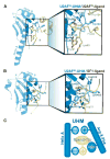
Structures of U2AF-UHM/ligand complexes. The UHM is shown in blue, the protein ligand is shown in yellow. Key interacting side chains are drawn in ball-and-stick representation. (A) The U2AF35-UHM bound to the U2AF65-ligand. (B) The U2AF65-UHM bound to the SF1-ligand. (C) Schematic representation of signature UHM protein–protein interactions shared by the two complexes.
Figure 4
Figure 4
Identification of candidate UHM-containing proteins. (A) Structure-based alignment of candidate UHM sequences, U2AF35-UHM, U2AF65-UHM, and representative canonical RRMs whose structures in complex with RNA are known. Structures were aligned using the program TOPP from the CCP4 suite (Collaborative Computational Project, no. 4, 1994). Secondary structure elements are indicated above the sequences. The positions of key residues that distinguish UHM from RRM domains are highlighted yellow. Consensus UHM residues including acidic residues and the Arg–X–Phe loop are red. Residues in the RNP-like motifs that match the canonical RRM consensus are blue. (B) Phylogenetic tree of candidate UHM domains.
Figure 5
Figure 5
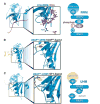
RNA recognition by an RRM domain compared with the U2AF-UHM domains. The UHM or RRM domains are shown in blue, protein ligands are yellow, and RNA ligands are purple. (A) The U1A-RRM recognizing an RNA oligonucleotide. The RNP1-Arg recognizes the RNA phosphates, a nucleotide base stacks on RNP2-Tyr, and RNP2-Leu is folded in the hydrophobic core. (B) The U2AF35-UHM/U2AF65-ligand complex (PDB code 1JMT) rotated 180° about the Y-axis with respect to Figure 3 to show the putative RNA interaction surface. (C) Similar view of the U2AF65-UHM/SF1-ligand structure (PDB code 1O0P). The RNP2-Ile packs with the ligand-Trp. The small aliphatic residues in the RNP-like motifs are predicted to be unable to form favorable RNA interactions. Schematic representation of RNP residues that differ between UHMs compared with canonical RRMs is shown to the right.

References

    1. Abovich N, Liao XC, Rosbash M. The yeast MUD2 protein: An interaction with PRP11 defines a bridge between commitment complexes and U2 snRNP addition. Genes & Dev. 1994;8:843–854. - PubMed
    1. Adam SA, Nakagawa T, Swanson MS, Woodruff TK, Dreyfuss G. mRNA polyadenylate-binding protein: Gene isolation and sequencing and identification of a ribo-nucleoprotein consensus sequence. Mol Cell Biol. 1986;6:2932–2943. - PMC - PubMed
    1. Allain FH, Bouvet P, Dieckmann T, Feigon J. Molecular basis of sequence-specific recognition of pre-ribosomal RNA by nucleolin. EMBO J. 2000;19:6870–6881. - PMC - PubMed
    1. Altschul SF, Madden TL, Schaffer AA, Zhang J, Zhang Z, Miller W, Lipman DJ. Gapped BLAST and PSI-BLAST: A new generation of protein database search programs. Nucleic Acids Res. 1997;25:3389–3402. - PMC - PubMed
    1. Anantharaman V, Koonin EV, Aravind L. Comparative genomics and evolution of proteins involved in RNA metabolism. Nucleic Acids Res. 2002;30:1427–1464. - PMC - PubMed

Publication types

LinkOut - more resources