. 2004 Jul 1;18(13):1630-42.
doi: 10.1101/gad.1194004.

Loss of C. elegans BBS-7 and BBS-8 protein function results in cilia defects and compromised intraflagellar transport

Affiliations

Loss of C. elegans BBS-7 and BBS-8 protein function results in cilia defects and compromised intraflagellar transport

Oliver E Blacque et al. Genes Dev. .

Abstract

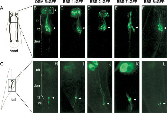
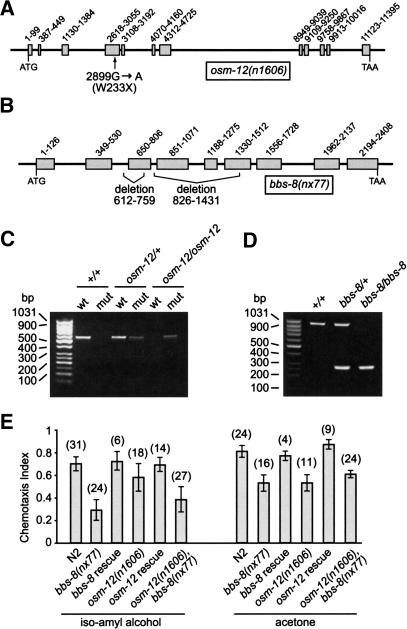
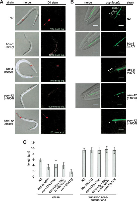
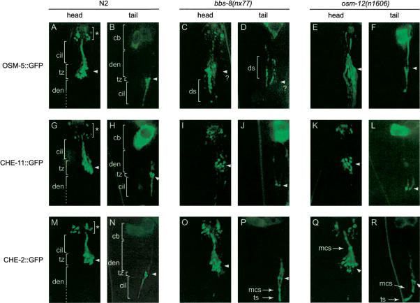
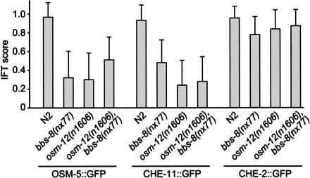
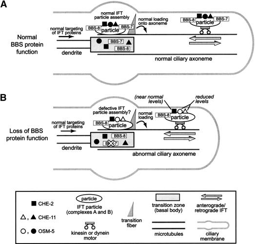
Bardet-Biedl syndrome (BBS) is a genetically heterogeneous developmental disorder whose molecular basis is largely unknown. Here, we show that mutations in the Caenorhabditis elegans bbs-7 and bbs-8 genes cause structural and functional defects in cilia. C. elegans BBS proteins localize predominantly at the base of cilia, and like proteins involved in intraflagellar transport (IFT), a process necessary for cilia biogenesis and maintenance, move bidirectionally along the ciliary axoneme. Importantly, we demonstrate that BBS-7 and BBS-8 are required for the normal localization/motility of the IFT proteins OSM-5/Polaris and CHE-11, and to a notably lesser extent, CHE-2. We propose that BBS proteins play important, selective roles in the assembly and/or function of IFT particle components. Our findings also suggest that some of the cardinal and secondary symptoms of BBS, such as obesity, diabetes, cardiomyopathy, and learning defects may result from cilia dysfunction.


Figures

Figure 1.
Figure 1.
C. elegans BBS proteins localize predominantly to the transition zones and axonemes of cilia. (A,G) Schematics of the head (A) and tail (G) indicate the position of cilial structures at the dendritic endings of amphid and phasmid neurons. (B–F,H–L) Fluorescent images of the head (amphids, B–F) and tail (phasmids, H–L) regions of N2 worms expressing GFP-tagged OSM-5 (B,H), BBS-1 (C,I), BBS-2 (D,J), BBS-7 (E,K), and BBS-8 (F,L). The BBS proteins localize specifically to the transition zones (C–F,I–L), and like the IFT protein, OSM-5 (B,H), they are also found along the ciliary axonemes. The ciliary axonemes (cil), transition zones (tz), dendrites (den), and cell bodies (cb) are indicated. An asterisk denotes the ciliated endings of neurons found at the anterior end of the worm (e.g., IL1/2, OLL, CEP, and BAG neurons). Arrowheads in all panels denote the amphid and phasmid transition zones. Note that in all panels, only one set of amphid and phasmid neurons is shown except for the schematic in A, where both sets (one on each side) are shown.
Figure 2.
Figure 2.
osm-12(n1606) and bbs-8(nx77) animals bearing mutations in the C. elegans bbs-7 and bbs-8 genes, respectively, exhibit chemotaxis defects. (A,B) Schematics depicting the G→A nonsense mutation at nucleotide 2899 of osm-12(n1606), and the position of two deletions in bbs-8(nx77). Exons are shown as shaded boxes. (C) Single worms containing the osm-12(n1606) nonsense mutation were identified using two PCR reactions that are designed to amplify wild-type (wt), osm-12(+), or mutant (mut) osm-12(n1606) genomic DNA fragments. Shown are the PCR products obtained from +/+, osm-12(n1606)/+, and osm-12(n1606)/osm-12(n1606) animals. (D) Single-worm PCR using primers that flank the 826–1431 deletion in bbs-8(nx77) identifies +/+, bbs-8(nx77)/+, and bbs-8(nx77)/bbs-8(nx77) animals. (E) The osm-12(n1606), bbs-8(nx77), and osm-12(n1606),bbs-8(nx77) mutant worms are defective in chemotaxis (Che) toward isoamyl alcohol and acetone. Rescue of this Che defect was achieved by expressing bbs-7(+) and bbs-8(+) genes in the osm-12(n1606) and bbs-8(nx77) mutant worms, respectively. The number of assays for each chemotaxis data point is indicated in brackets. Error bars, ±S.E.M.
Figure 3.
Figure 3.
osm-12(n1606) and bbs-8(nx77) mutants possess disrupted ciliary structures. (A) osm-12 and bbs-8 animals are severely defective in the ability to incorporate the dye, DiI, into their amphid and phasmid neurons via ciliated endings. The dye-uptake defective (Dyf) phenotype was fully rescued by expressing bbs-7(+) and bbs-8(+) genes in the osm-12 and bbs-8 worms, respectively. Note that barely detectable DiI staining in both bbs gene mutants was observed only after prolonged exposure (exp.) times. (B,C) Using the ASER neuron-specific marker, gcy-5p::gfp, the ASER cilium structure of osm-12(n1606), bbs-8(nx77), and osm-12(n1606),bbs-8(nx77) mutants is observed to be shorter than that of N2 controls. The ciliary axoneme (cil), transition zone (tz), and dendrite (den) structures of ASER are indicated at top, right. An asterisk also denotes the position of the transition zone in each panel. Also shown is the fragmented GFP staining pattern (see arrowheads) that is often observed in the bbs mutants. The cilium morphology data presented in C shows that the ASER cilium structure of osm-12, bbs-8, and osm-12,bbs-8 mutants is significantly shorter (P < 0.05) than N2 controls, but not as short as the ASER cilium in osm-5(p813) mutant animals. Also shown is the identical relative positioning of the ASER transition zone (tz) in the bbs gene mutants compared with N2 worms (measured as tz → anterior end of worm, seen in the DIC–GFP merged images). Each data point represents measurements from 50 different animals. Error bars, ±S.E.M. Bars, 5 μm.
Figure 4.
Figure 4.
BBS-7 and BBS-8 protein function is required for the proper localization of the GFP-tagged IFT particle components OSM-5 and CHE-11, and to a lesser degree CHE-2. Shown are representative fluorescent images of the anterior (amphids) and posterior (phasmids) ends of N2, bbs-8(nx77) and osm-12(n1606) worms expressing the identical osm-5::gfp, che-11::gfp, and che-2::gfp transgenes. In contrast to the localization of GFP-tagged OSM-5 and CHE-11 to the ciliary axoneme in N2 worms (A,B,G,H), these two IFT proteins generally display abnormal staining along the ciliary axonemes of bbs-8(nx77) (C,D,I,J) and osm-12(n1606) (E,F,K,L) mutant animals. In contrast, there is strong localization of CHE-2::GFP to the ciliary axonemes of the bbs-8(nx77) (O,P) and osm-12(n1606) (Q,R) mutants, similar to that seen in N2 control worms (M,N). The ciliary axonemes (cil), transition zone (tz), and dendrite (den) are indicated in each panel. In some of the bbs gene mutants, spots of GFP fluorescence are often observed in the dendrites (ds), near the midpoint of the ciliary axoneme (mcs) and at the distal end of the ciliary axoneme (tip spot, or ts). Note that only one set of amphid and phasmid cilia are shown in each panel. A question mark denotes the difficulty in precisely identifying the location of the transition zones in panels C and D.
Figure 5.
Figure 5.
The C. elegans BBS-7 and BBS-8 proteins are required for efficient IFT. The IFT motility of OSM-5 and CHE-11, but not CHE-2 is significantly compromised in the absence of BBS-7 and BBS-8 protein function. The osm-5::gfp, che-11::gfp, and che-2::gfp transgenes were crossed from wild-type (N2) to each of the bbs single or double mutants (bbs-8(nx77), osm-12(n1606), and osm-12(n1606),bbs-8(nx77) and the amphid (n = 20) and phasmid (n = 20) IFT motilities were recorded in movies (see Materials and Methods). In a blind assay, IFT was scored on a discontinuous scale where 0 equals no detectable IFT, 0.25 equals barely detectable IFT (i.e., extremely weak signals indicating moving particles that are sometimes observed), 0.50 equals weak IFT (i.e., faint signals of continuous particle movement in both the anterograde and retrograde directions), 0.75 equals moderate levels of IFT, and 1 equals IFT that is highly prominent and operating in both the anterograde and retrograde directions. No significant difference between amphid and phasmid IFT was detected; therefore, the scores above are a combination of the amphid and phasmid IFT scores (i.e., n = 40). Error bars, S.E.M.
Figure 6.
Figure 6.
Model of BBS-7 and BBS-8 protein function. (A) In wild-type cilia, BBS-7 and BBS-8 proteins, as well as complex A and B IFT particle components (e.g., OSM-5, CHE-11 and CHE-2) are targeted to, and accumulate at the base of the cilium structure. Within the basal body region (including the transitional fibers), the BBS-7 and BBS-8 proteins may play a role in facilitating the selective assembly of IFT protein components into the IFT particle, perhaps by acting as molecular chaperones or directly associating with them. Fully assembled motor–IFT particle–cargo complexes, which likely include the BBS proteins, are subsequently loaded onto the axoneme proper, where they undergo IFT. Note that for simplicity, only one transitional fiber is shown. (B) In cilia where BBS-7 or BBS-8 protein function is lost (shown only for BBS-7), the IFT components are targeted normally to the base of cilia. However, IFT particle assembly within the basal body region may be disrupted, resulting in the failure of certain IFT protein components (e.g., OSM-5 and CHE-11, but not CHE-2) to be efficiently incorporated into the final IFT particle complex and, hence, undergo IFT. This defect results in compromised IFT and ciliary defects (e.g., short and/or structurally abnormal cilia).

Similar articles

Cited by

References

    1. Ansley S.J., Badano, J.L., Blacque, O.E., Hill, J., Hoskins, B.E., Leitch, C.C., Kim, J.C., Ross, A.J., Eichers, E.R., Teslovich, T.M., et al. 2003. Basal body dysfunction is a likely cause of pleiotropic Bardet-Biedl syndrome. Nature 425: 628-633. - PubMed
    1. Badano J.L., Ansley, S.J., Leitch, C.C., Lewis, R.A., Lupski, J.R., and Katsanis, N. 2003. Identification of a novel Bardet-Biedl syndrome protein, BBS7, that shares structural features with BBS1 and BBS2. Am. J. Hum. Genet. 72: 650-658. - PMC - PubMed
    1. Bale A.E. 2002. Hedgehog signaling and human disease. Annu. Rev. Genomics Hum. Genet. 3: 47-65. - PubMed
    1. Bargmann C.I., Hartwieg, E., and Horvitz, H.R. 1993. Odorant-selective genes and neurons mediate olfaction in C. elegans. Cell 74: 515-527. - PubMed
    1. Blatch G.L. and Lassle, M. 1999. The tetratricopeptide repeat: A structural motif mediating protein–protein interactions. Bioessays 21: 932-939. - PubMed

Publication types

MeSH terms