. 2004 Jul;186(14):4585-95.
doi: 10.1128/JB.186.14.4585-4595.2004.

Bacillus subtilis YdiH is a direct negative regulator of the cydABCD operon

Affiliations

Bacillus subtilis YdiH is a direct negative regulator of the cydABCD operon

Matthew Schau et al. J Bacteriol. 2004 Jul.

Abstract

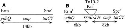
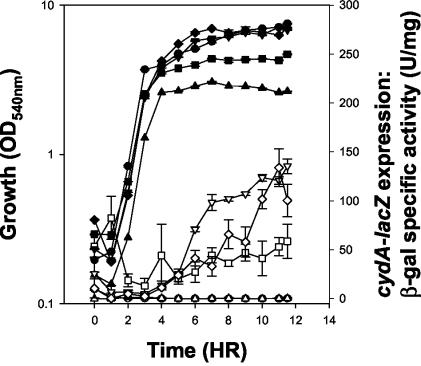
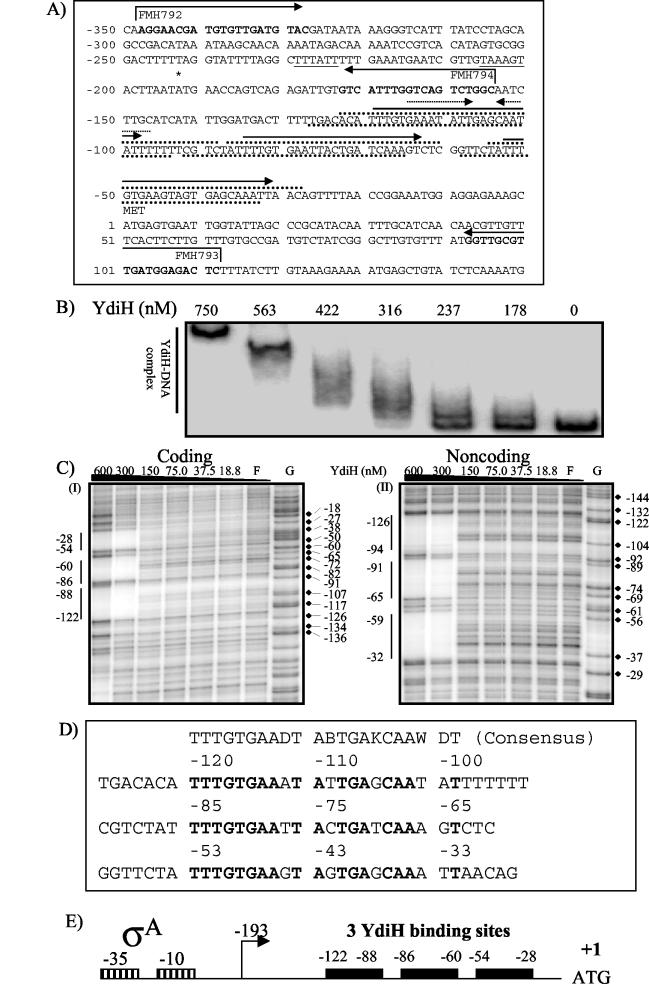
During aerobic respiration, Bacillus subtilis utilizes three terminal oxidases, cytochromes aa3, caa3, and bd. Cytochrome bd is encoded by the cydABCD operon. We report here the first identification of a regulator for the cydABCD operon, YdiH. While working with DeltaresDE mutant strains, we identified colonies which contained suppressor mutations (cmp) which bypassed the requirement for ResD for all phenotypes not associated with cytochrome aa3 or caa3. Mapping identified a class of Tn10 insertions which were close to the cmp locus (Tn10-2) and a second class (Tn10-1) which was inserted in cydD, a gene which appears to be essential to the cmp phenotype. Sequencing of the cmp loci from four independent DeltaresDE cmp isolates yielded four loss-of-function alleles of ydiH, a gene encoding a protein with homology to AT-rich DNA-binding proteins. Additionally, we determined that cytochrome bd was aberrantly expressed in the DeltaresDE cmp background. Together these data led to the hypothesis that YdiH serves as a negative regulator of cydABCD expression, a hypothesis supported by both gel-shift and DNase I footprinting analyses. YdiH protected the cydA promoter region at three 22-bp repeats located in the long 5' untranslated region (193 bp). Induction of the cydABCD operon in a DeltaresDE background showed that expression of the terminal oxidase bd was responsible for the bypass phenotype observed in a DeltaresDE cmp strain, indicating that cytochrome bd expression complemented the loss of cytochromes aa3 and caa3 in the DeltaresDE strain.


Figures

FIG. 1.
FIG. 1.
Transformation mapping of the cmp locus. The frequency of each transformant obtained is shown in Table 4. Three-factor cross transformation using MH5881 (ΔydhQ ΔtatCY) as the donor strain and MH5857 (cmp) as the recipient strain was selected on Spcr and screened for Ermr and the cmp phenotype, as shown in Table 4. Three-factor cross transformation using MH5874 (ΔtatCY rrnE-23S::Tn10-2) as the donor strain and MH5857 (cmp) was selected on Spcr and screened for Neor and for the cmp phenotype, as shown in Table 4. (A) The first three-factor cross (cross 1 in Table 4) places cmp in the 14-kb region between ydhQ and tatCY. (B) The second three-factor cross (cross 2 in Table 4) localizes cmp between the Tn10-2 insertion and tatCY.
FIG. 2.
FIG. 2.
Growth and cydA-lacZ expression from B. subtilis strains cultured for 11.5 h in LB medium supplemented with 0.5% glucose. Growth, solid symbols; β-galactosidase, open symbols. •, MH5878 (wild-type cydA-lacZ); ▴, MH5880 (ΔresDE cydA-lacZ); ▾, MH5879 (ΔresDE cmp cydA-lacZ); ▪, MH5891 (ΩydiH cydA-lacZ); ♦, MH5893 (ΔresDE ΩydiH cydA-lacZ).
FIG. 3.
FIG. 3.
Light absorption difference (dithionite reduced minus ferricyanide oxidized) spectra of membranes from strains JH642 (wild type), MH5202 (ΔresDE), MH5887 (ΔresDE cmp), MH5891 (ΩydiH), and MH5893 (ΩydiH ΔresDE). B. subtilis strains were grown in LB medium with 0.5% glucose and harvested during stationary phase. A representative spectrum is shown for each strain. (A) JH642. (B) MH5857. (C) MH5202. (D) MH5891. (E) MH5893.
FIG. 4.
FIG. 4.
YdiH binds directly to the cydA promoter. (A) Structure of the cydA regulatory region. The major transcriptional start site is shown with an asterisk. The inverted repeats and tandem repeats are indicated as dotted line arrows or solid arrows, respectively. The YdiH protected regions revealed by the DNase I footprinting assay are shown by dotted lines above (coding strand) and below (noncoding strand) the sequence. The consensus sequences for the −35 and −10 σA recognition sequences are underlined. The primer sequences used to generate PCR products are in boldface, and broken arrows indicate the 5′-to-3′ sequence. The numbering is relative to the translation start (ATG) as +1 (also for panels C and D). (B) Gel shift assay of the cydA promoter with YdiH. The probe was the PCR product using radiolabeled primers FMH792 and FMH793 and JH642 chromosomal DNA as template. The primer was labeled as described in Materials and Methods. The concentrations of YdiH used are indicated at the top of each lane. (C) DNase I footprinting of the cydA promoter by using YdiH. Labeled DNA fragments were generated as in panel B. For the coding or noncoding footprint, FMH792 or FMH793 was end labeled, respectively. The YdiH concentrations are shown at the top of each lane. F, free of YdiH; G, Maxam-Gilbert G-sequencing reaction lane as a marker. The vertical lines show the YdiH protected regions. (D) The cydA promoter alignment for directed repeats. The consensus sequence is shown above, and the IUPAC (the International Union of Pure and Applied Chemistry) ambiguity codes for DNA were used: D = A, T, or G; B = T, C, or G; K = G or T; and W = A or T. The identical base pairs among the three repeats are in bold. (E) Graphic representation of YdiH binding on the cydA promoter. Solid black boxes represent the three YdiH binding site on the cydA promoter region. The boxes with vertical lines represent the consensus −10 and −35 for σA identified previously (35). The numbering is relative to the translational start (ATG) as +1. For simplicity, only coding strand protected regions are numbered. The transcription start site is identified with a broken arrow.

Similar articles

Cited by

References

    1. Bisschop, A., and W. N. Konings. 1976. Reconstitution of reduced nicotinamide adenine dinucleotide oxidase activity with menadione in membrane vesicles from the menaquinone-deficient Bacillus subtilis aroD. Relation between electron transfer and active transport. Eur. J. Biochem. 67:357-365. - PubMed
    1. Carls, R. A., and R. S. Hanson. 1971. Isolation and characterization of tricarboxylic acid cycle mutants of Bacillus subtilis. J. Bacteriol. 106:848-855. - PMC - PubMed
    1. Chary, V. K., E. I. Amaya, and P. J. Piggot. 1997. Neomycin- and spectinomycin-resistance replacement vectors for Bacillus subtilis. FEMS Microbiol. Lett. 153:135-139. - PubMed
    1. Cutting, S. M., and P. B. Vander Horn. 1990. Genetic analysis, p. 24-74. In C. R. Harwood and S. M. Cutting (ed.), Molecular biological methods for Bacillus. John Wiley & Sons, Inc., New York, N.Y.
    1. de Vrij, W., B. van den Burg, and W. N. Konings. 1987. Spectral and potentiometric analysis of cytochromes from Bacillus subtilis. Eur. J. Biochem. 166:589-595. - PubMed

Publication types

MeSH terms

LinkOut - more resources