. 2004 Jul;186(14):4645-54.
doi: 10.1128/JB.186.14.4645-4654.2004.

Structure and electrophysiological properties of the YscC secretin from the type III secretion system of Yersinia enterocolitica

Affiliations

Structure and electrophysiological properties of the YscC secretin from the type III secretion system of Yersinia enterocolitica

Peter Burghout et al. J Bacteriol. 2004 Jul.

Abstract

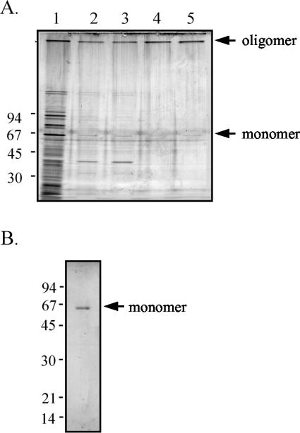
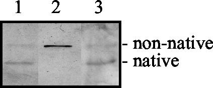
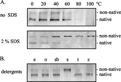
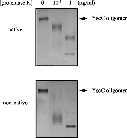
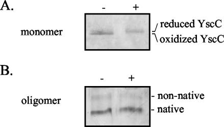
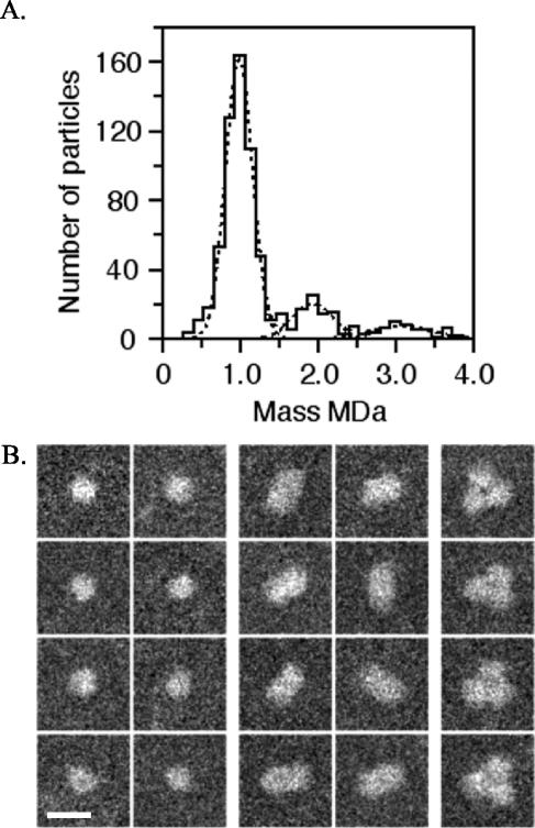
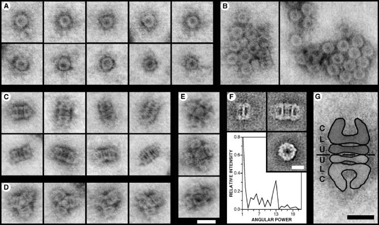
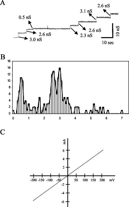
YscC is the integral outer membrane component of the type III protein secretion machinery of Yersinia enterocolitica and belongs to the family of secretins. This group of proteins forms stable ring-like oligomers in the outer membrane, which are thought to function as transport channels for macromolecules. The YscC oligomer was purified after solubilization from the membrane with a nonionic detergent. Sodium dodecyl sulfate did not dissociate the oligomer, but it caused a change in electrophoretic mobility and an increase in protease susceptibility, indicating partial denaturation of the subunits within the oligomer. The mass of the homo-oligomer, as determined by scanning transmission electron microscopy, was approximately 1 MDa. Analysis of the angular power spectrum from averaged top views of negatively stained YscC oligomers revealed a 13-fold angular order, suggesting that the oligomer consists of 13 subunits. Reconstituted in planar lipid bilayers, the YscC oligomer displayed a constant voltage-independent conductance of approximately 3 nS, thus forming a stable pore. However, in vivo, the expression of YscC did not lead to an increased permeability of the outer membrane. Electron microscopy revealed that the YscC oligomer is composed of three domains, two stacked rings attached to a conical domain. This structure is consistent with the notion that the secretin forms the upper part of the basal body of the needle structure of the type III secreton.


Figures

FIG. 1.
FIG. 1.
Purification of the YscC oligomer with the nonionic detergent Elugent. (A) Protein fractions of the different steps of the purification procedure were analyzed on a 3 to 15% polyacrylamide gradient gel and stained with silver. Samples were adjusted to contain equal amounts of YscC oligomer. Lane 1, protein fraction from whole cells (lane 1); lane 2, protein fraction from cell envelopes; lane 3, soluble protein fraction obtained after membrane extraction with Elugent; lane 4, protein fraction obtained after sucrose gradient centrifugation; lane 5, purified YscC oligomer after ion-exchange chromatography. (B) The purified YscC oligomer was dissociated with hot phenol, loaded on an 11% polyacrylamide gel, and visualized with Coomassie brilliant blue staining. The positions of the molecular mass markers (in kilodaltons) and of the YscC oligomer and monomer are indicated to the left and right of the gels, respectively.
FIG. 2.
FIG. 2.
Effects of heat and SDS on the purified YscC oligomer. The seminative 3 to 9% polyacrylamide gradient gel was loaded with purified YscC oligomer (lane 1), purified YscC oligomer that had been incubated for 10 min at 100°C in the presence of 2% SDS (lane 2), and cell envelopes from the pYV-cured Y. enterocolitica strain CE1525 carrying plasmids pSM3km and pRS6. Proteins were visualized by staining with silver. The positions of the native and partially denatured (nonnative) forms of the YscC oligomer are indicated to the right of the gel.
FIG. 3.
FIG. 3.
Stability of the native YscC oligomer. (A) Elugent-solubilized YscC oligomer was incubated in the absence or presence of 2.0% SDS for 10 min at the indicated temperatures. (B) Elugent-solubilized YscC oligomer was incubated in the presence of 2% Elugent (e), OPOE (o), SB12 (sb), SDS (s), Triton X-100 (t), or Zwittergent 3-14 (z) for 1 h at 40°C. All samples were loaded on seminative 3 to 9% polyacrylamide gradient gels, subjected to electrophoresis, and stained with silver. The positions of the native and nonnative forms of the YscC oligomer are indicated to the right of the gels.
FIG. 4.
FIG. 4.
Protease susceptibility of the purified YscC oligomer. Purified YscC oligomers were incubated with 2% SDS for 10 min at room temperature for analysis of the native form or at 100°C for analysis of the nonnative form. The oligomers were then incubated with the indicated concentrations of proteinase K for 10 min at room temperature. Samples were loaded on a 3 to 9% polyacrylamide gradient gel. The position of the YscC oligomer to indicated on the right of the gels. Proteins were visualized by staining with silver.
FIG. 5.
FIG. 5.
Role of disulfide bonds in the stability of the SDS-sensitive YscC oligomer. Purified YscC oligomer was incubated in the absence (−) or presence (+) of 20 mM DTT for 10 min at 40°C and was either dissociated into monomers by treatment with TCA and TFA and loaded on a 8% polyacrylamide gel (A) or directly loaded on a seminative 3 to 9% polyacrylamide gradient gel (B). The positions of the reduced and oxidized forms of the YscC monomer and the native and nonnative forms of the YscC oligomer are indicated to the right of the gels. Proteins were visualized by staining with silver.
FIG. 6.
FIG. 6.
STEM mass analysis of unstained, Elugent-purified YscC oligomers. (A) Histogram of the mass values. The different populations have masses of 985 ± 175 kDa (n = 546), 1,928 ± 254 kDa (n = 97), and 3,079 ± 374 kDa (n = 53), indicating the presence of one, two, and three YscC oligomers, respectively. Correction was made for beam-induced mass loss using the factor 1.029 (see Materials and Methods). (B) Galleries showing particles with masses in the range of the first, second, and third mass peaks (left, middle, and right galleries, respectively). Protein is displayed in white. Bar, 25 nm.
FIG. 7.
FIG. 7.
STEM analysis of Elugent-purified YscC oligomers. (A to E) STEM images of YscC oligomers that have been negatively stained with uranyl acetate. The contrast of the dark-field images has been inverted to show protein as gray. (A) Ring-like top views. (B) Rosette-shaped aggregates showing predominantly top views of the oligomer. (C) Rectangular side views that correspond to dimers of the oligomer. The thick band at the center of the oligomer resolves into two in some projections. The outer ends of the oligomer display various orientations. (D) Star-shaped side views of trimeric aggregates. (E) Essentially square side views formed by the association of four or five oligomers. The micrographs in panels A to E are all shown at the same magnification. Bar, 20 nm. (F) TEM analysis. (Top left) Average side-view projection of a single oligomer (n = 8 of 16); (top right) average side-view projection of two oligomers associated end-to-end (n = 16 of 16); (bottom right) average top view projection (n = 58 of 81). Bar, 10 nm. The graph shows the angular power spectrum of the top-view average, illustrating the presence of a strong 13-fold angular harmonic. (G) Interpretation of the rectangular side views demonstrated using a STEM image. The protein structure is highlighted, and a discontinuous black line indicates the interface between the two oligomers. The different domains within the structure, i.e., the conical domain (C), the lower ring (L), and the upper ring (U), are labeled. Bar, 10 nm.
FIG. 8.
FIG. 8.
Pore activity of the YscC oligomer. (A) Channel recording of multiple insertions of the purified YscC oligomer in a planar lipid bilayer at a membrane potential of 100 mV. The sizes of the transitions are indicated. (B) Amplitude histogram of channel openings (n = 204) at 100 mV. (C) Voltage-ramp analysis of multiple YscC channels from 0 to 200 mV and 0 to −200 mV over a total time span of 200 s.

Similar articles

Cited by

References

    1. Allaoui, A., R. Scheen, C. Lambert de Rouvroit, and G. R. Cornelis. 1995. VirG, a Yersinia enterocolitica lipoprotein involved in Ca2+ dependency, is related to exsB of Pseudomonas aeruginosa. J. Bacteriol. 177:4230-4237. - PMC - PubMed
    1. Bitter, W., M. Koster, M. Latijnhouwers, H. de Cock, and J. Tommassen. 1998. Formation of oligomeric rings by XcpQ and PilQ, which are involved in protein transport across the outer membrane of Pseudomonas aeruginosa. Mol. Microbiol. 27:209-219. - PubMed
    1. Blocker, A., N. Jouihri, E. Larquet, P. Gounon, F. Ebel, C. Parsot, P. Sansonetti, and A. Allaoui. 2001. Structure and composition of the Shigella flexneri “needle complex,” a part of its type III secreton. Mol. Microbiol. 39:652-663. - PubMed
    1. Brok, R., P. Van Gelder, M. Winterhalter, U. Ziese, A. J. Koster, H. de Cock, M. Koster, J. Tommassen, and W. Bitter. 1999. The C-terminal domain of the Pseudomonas secretin XcpQ forms oligomeric rings with pore activity. J. Mol. Biol. 294:1169-1179. - PubMed
    1. Burghout, P., F. Beckers, E. de Wit, R. van Boxtel, G. R. Cornelis, J. Tommassen, and M. Koster. Role of the pilot protein YscW in the biogenesis of the secretin YscC in Yersinia enterocolitica. J. Bacteriol., in press. - PMC - PubMed

Publication types

MeSH terms

LinkOut - more resources