

Evidence that the algI/algJ gene cassette, required for O acetylation of Pseudomonas aeruginosa alginate, evolved by lateral gene transfer
- PMID: 15231808
- PMCID: PMC438637
- DOI: 10.1128/JB.186.14.4759-4773.2004
Evidence that the algI/algJ gene cassette, required for O acetylation of Pseudomonas aeruginosa alginate, evolved by lateral gene transfer
Abstract
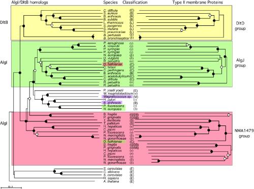
Pseudomonas aeruginosa strains, isolated from chronically infected patients with cystic fibrosis, produce the O-acetylated extracellular polysaccharide, alginate, giving these strains a mucoid phenotype. O acetylation of alginate plays an important role in the ability of mucoid P. aeruginosa to form biofilms and to resist complement-mediated phagocytosis. The O-acetylation process is complex, requiring a protein with seven transmembrane domains (AlgI), a type II membrane protein (AlgJ), and a periplasmic protein (AlgF). The cellular localization of these proteins suggests a model wherein alginate is modified at the polymer level after the transport of O-acetyl groups to the periplasm. Here, we demonstrate that this mechanism for polysaccharide esterification may be common among bacteria, since AlgI homologs linked to type II membrane proteins are found in a variety of gram-positive and gram-negative bacteria. In some cases, genes for these homologs have been incorporated into polysaccharide biosynthetic operons other than for alginate biosynthesis. The phylogenies of AlgI do not correlate with the phylogeny of the host bacteria, based on 16S rRNA analysis. The algI homologs and the gene for their adjacent type II membrane protein present a mosaic pattern of gene arrangement, suggesting that individual components of the multigene cassette, as well as the entire cassette, evolved by lateral gene transfer. AlgJ and the other type II membrane proteins, although more diverged than AlgI, contain conserved motifs, including a motif surrounding a highly conserved histidine residue, which is required for alginate O-acetylation activity by AlgJ. The AlgI homologs also contain an ordered series of motifs that included conserved amino acid residues in the cytoplasmic domain CD-4; the transmembrane domains TM-C, TM-D, and TM-E; and the periplasmic domain PD-3. Site-directed mutagenesis studies were used to identify amino acids important for alginate O-acetylation activity, including those likely required for (i) the interaction of AlgI with the O-acetyl precursor in the cytoplasm, (ii) the export of the O-acetyl group across the cytoplasmic membrane, and (iii) the transfer of the O-acetyl group to a periplasmic protein or to alginate. These results indicate that AlgI belongs to a family of membrane proteins required for modification of polysaccharides and that a mechanism requiring an AlgI homolog and a type II membrane protein has evolved by lateral gene transfer for the esterification of many bacterial extracellular polysaccharides.
Copyright 2004 American Society for Microbiology
Figures


Similar articles
-
Mutant analysis and cellular localization of the AlgI, AlgJ, and AlgF proteins required for O acetylation of alginate in Pseudomonas aeruginosa.J Bacteriol. 2002 Jun;184(11):3000-7. doi: 10.1128/JB.184.11.3000-3007.2002. J Bacteriol. 2002. PMID: 12003941 Free PMC article.
-
Identification of algI and algJ in the Pseudomonas aeruginosa alginate biosynthetic gene cluster which are required for alginate O acetylation.J Bacteriol. 1996 Apr;178(8):2186-95. doi: 10.1128/jb.178.8.2186-2195.1996. J Bacteriol. 1996. PMID: 8636017 Free PMC article.
-
Analysis of the alginate O-acetylation machinery in Pseudomonas aeruginosa.Appl Microbiol Biotechnol. 2020 Mar;104(5):2179-2191. doi: 10.1007/s00253-019-10310-6. Epub 2020 Jan 4. Appl Microbiol Biotechnol. 2020. PMID: 31900562
-
Pseudomonas aeruginosa biofilms: role of the alginate exopolysaccharide.J Ind Microbiol. 1995 Sep;15(3):162-8. doi: 10.1007/BF01569821. J Ind Microbiol. 1995. PMID: 8519473 Review.
-
Proteolytic regulation of alginate overproduction in Pseudomonas aeruginosa.Mol Microbiol. 2012 May;84(4):595-607. doi: 10.1111/j.1365-2958.2012.08049.x. Epub 2012 Apr 13. Mol Microbiol. 2012. PMID: 22497280 Free PMC article. Review.
Cited by
-
In silico detection of virulence gene homologues in the human pathogen sphingomonas spp.Evol Bioinform Online. 2014 Dec 11;10:229-38. doi: 10.4137/EBO.S20710. eCollection 2014. Evol Bioinform Online. 2014. PMID: 25574122 Free PMC article.
-
The mechanism of peptidoglycan O-acetylation in Gram-negative bacteria typifies bacterial MBOAT-SGNH acyltransferases.bioRxiv [Preprint]. 2024 Sep 19:2024.09.17.613324. doi: 10.1101/2024.09.17.613324. bioRxiv. 2024. Update in: J Biol Chem. 2025 Jun;301(6):108531. doi: 10.1016/j.jbc.2025.108531. PMID: 39345430 Free PMC article. Updated. Preprint.
-
ClpXP proteases positively regulate alginate overexpression and mucoid conversion in Pseudomonas aeruginosa.Microbiology (Reading). 2008 Jul;154(Pt 7):2119-2130. doi: 10.1099/mic.0.2008/017368-0. Microbiology (Reading). 2008. PMID: 18599839 Free PMC article.
-
Acyltransferases that Modify Cell Surface Polymers Across the Membrane.Biochemistry. 2025 Apr 15;64(8):1728-1749. doi: 10.1021/acs.biochem.4c00731. Epub 2025 Apr 2. Biochemistry. 2025. PMID: 40171682 Review.
-
Biosynthesis of the Pseudomonas aeruginosa Extracellular Polysaccharides, Alginate, Pel, and Psl.Front Microbiol. 2011 Aug 22;2:167. doi: 10.3389/fmicb.2011.00167. eCollection 2011. Front Microbiol. 2011. PMID: 21991261 Free PMC article.
References
-
- Altschul, S. F., W. Gish, W. Miller, E. W. Myers, and D. J. Lipman. 1990. Basic local alignment search tool. J. Mol. Biol. 215:403-410. - PubMed
-
- Ausubel, F. M., R. Brent, R. E. Kingston, D. D. Moore, J. G. Seidman, J. A. Smith, and K. Struhl (ed.). 1993. Current protocols in molecular biology, vol. 2. Greene Publishing Associates, Inc./John Wiley & Sons, Inc., New York, N.Y.
-
- Baltimore, R. S., and M. Mitchell. 1982. Immunologic investigations of mucoid strains of Pseudomonas aeruginosa: comparison of susceptibility to opsonic antibody in mucoid and nonmucoid strains. J. Infect. Dis. 141:238-247. - PubMed
Publication types
MeSH terms
Substances
Grants and funding
LinkOut - more resources
Full Text Sources
Other Literature Sources
Molecular Biology Databases
Research Materials
