. 2004 Oct 1;279(40):42240-9.
doi: 10.1074/jbc.M406775200. Epub 2004 Jul 1.

BAD is a pro-survival factor prior to activation of its pro-apoptotic function

Affiliations

BAD is a pro-survival factor prior to activation of its pro-apoptotic function

So Young Seo et al. J Biol Chem. .

Abstract

The mammalian BAD protein belongs to the BH3-only subgroup of the BCL-2 family. In contrast to its known pro-apoptotic function, we found that endogenous and overexpressed BAD(L) can inhibit cell death in neurons and other cell types. Several mechanisms regulate the conversion of BAD from an anti-death to a pro-death factor, including alternative splicing that produces the N-terminally truncated BAD(S). In addition, caspases convert BAD(L) into a pro-death fragment that resembles the short splice variant. The caspase site that is selectively cleaved during cell death following growth factor (interleukin-3) withdrawal is conserved between human and murine BAD. A second cleavage site that is required for murine BAD to promote death following Sindbis virus infection, gamma-irradiation, and staurosporine treatment is not conserved in human BAD, consistent with the inability of human BAD to promote death with these stimuli. However, loss of the BAD N terminus by any mechanism is not always sufficient to activate its pro-death activity, suggesting that the N terminus is a regulatory domain rather than an anti-death domain. These findings suggest that BAD is more than an inert death factor in healthy cells; it is also a pro-survival factor, prior to its role in promoting cell death.


Figures

Fig. 1
Fig. 1. Cell type and age-dependent regulation of cell death by mBAD
A, cell death of COS-1 cells was determined by trypan blue staining at 24 h after infection with the indicated recombinant SV encoding HA-tagged murine BADL or HA-tagged human BCL-xL proteins (mean ± S.E. of three independent experiments). B, neuronal death in tissue slices cultured for 3 days prior to infection was determined by fluorescence-based computer imaging of PI staining at 68 h after infection with recombinant viruses expressing mBADL or mBIMS (mean ± S.E. of three independent experiments). C, cell death of 2-day-old cultures of dissociated cortical neurons prepared from individual bad+/+, bad+/−, and bad−/− embryos was determined by counting >3000 dual stained (PI-Hoechst) cells in 15 randomly selected fields per sample at 0 and 24 h after infection with Sindbis virus (SV-control) and subsequently correlated to genotypes. Neuronal death in 2-day-old cultures of spinal cord slices prepared from genotyped newborn littermates was determined as described for B (bad+/− cultures were not tested). Data shown are the mean ± S.E. for three independent experiments (*, p < 0.05 compared with bad−/−). D, immunoblot of lysates from neuronal cultures described in C or brain tissue from bad+/+ (Wt) and bad−/− (KO) mice using anti-BAD antibody C-20 (Santa Cruz Biotechnology). E, percentage of mortality following intracranial injection of the indicated recombinant Sindbis viruses in 3-day-old CD-1 mice (n > 65/group) or 5-week-old C57BL/6 mice (n > 30/group). F, cell death of rat embryonic cortical neurons was determined at 48 h after infection with the indicated recombinant viruses as described for C (counting > 1000 cells/sample in each of three independent experiments). An immunoblot for BAD in SV-control (SV-C) and SV-mBADL (mB)-infected cortical neuron cultures is shown (lower panel). G, representative fields of PI-Hoechst-stained cortical neurons from F are shown. H, summary of the age-dependent function of BAD. The precise neuron population responsible for virus-induced mortality is not known but is likely to involve the brain stem that regulates vital functions.
Fig. 2
Fig. 2. Caspase cleavage of mBAD promotes death in mature neurons
A and B, in vitro translated 35S-labeled wild type mBADL and derived point mutants were treated with buffer only, caspase-3 or -7 and analyzed by SDS-PAGE and autoradiography. (A potential unidentified caspase-7 cleavage product is marked with a dash.). C, cell death of infected rat cortical neurons was determined as described for Fig. 1, F and G. D, mortality of 5-week-old C57BL/6 mice (n > 20/group from three independent experiments) was determined as described for Fig. 1E. E, viability was determined by morphology of GFP-positive cells at 48 h after cotransfection of 6–7-day-old rat cortical neuron cultures with the indicated plasmids (5 μg) plus GFP (1 μg) in the absence or presence of 150 nm staurosporine (STR) during the final 24 h. F, summary of the role of caspases in converting BAD into a pro-death factor in mature neurons.
Fig. 3
Fig. 3. Uncleavable mBAD protects stable cell lines from apoptotic stimuli
A, immunoblot analysis of the stably transfected CCL10-BHK cells expressing the indicated HA-tagged constructs (individual cell clones are numbered, 1 and 2), loading 20–25 μg/lane and blotting with anti-BAD antibody C-20 (Santa Cruz Bio-technology). B, death of the cells shown in A was determined by trypan blue staining at 48 h after exposure to the indicated doses of γ-irradiation (mean ± S.E. for three independent experiments counting > 150 cells/sample). C, immunoblot analysis of the indicated stable CCL10-BHK cells with or without treatment with 1 μm staurosporine (STR) for 3 h was performed as described for A. Arrows mark full-length and cleaved BAD. D, immunoblot analysis (anti-BAD C-20) of individual BHK-21 cell clones (numbered, 1 and 2) stably transfected with the indicated constructs. E and F, cell death of BHK-21 cell line from D was determined by PI staining, flow cytometry, and/or manual counting at 24 h after infection with control Sindbis virus or 3 h after treatment with 1 μm staurosporine. Data presented are the mean ± S.E. from duplicate determinations in each of three independent experiments.
Fig. 4
Fig. 4. Murine BAD enhances cell death, whereas uncleavable BAD suppresses cell death in transient assays
A, percentage of death of HEK293 cells at 48 h after transfection of the indicated plasmids was determined by morphology. B, percentage of cell death of COS-1 cells was determined at 48 h after infection with the indicated recombinant Sindbis viruses. Data were compiled from three independent experiments that were analyzed by PI staining and flow cytometry or by trypan blue staining counting >200 cells/sample. C, immunoblot of BHK cell lysates at 24 h after infection using anti-BAD antibody C-20 (Santa Cruz Biotechnology).
Fig. 5
Fig. 5. Regulation of human BAD function by splicing
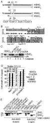
A, diagram of the long and short mouse and human BAD proteins produced by alternative splicing. Caspase cleavage site positions are numbered. B, alignment of predicted N-terminal amino acid sequences of BAD from mouse (mus), rat (rat), cow (bos) (Bos taurus, BF043486), pig (pig) (Sus scrofa, BF441436), and human (hum) chromosome 11 (hChr11). Expressed sequence tags for human slice variants 2 (NM_032989) and 1 (NM_004322) also lack the long N terminus. The predicted N termini of long and short splice variants (arrowheads) and potential caspase cleavage sites (thick vertical lines with amino acid numbers) are indicated. C, autoradiograph of in vitro translated (untagged) hBADS and derived point mutants as described for Fig. 2A. D, cell death of the indicated stably transfected CCL10-BHK cell clones (numbered) following exposure to the indicated doses of γ-irradiation was determined as described for Fig. 3B. (No cells transfected with wild type human BADS survived selection.) An immunoblot of untreated cell extracts is shown. E, immunoblot of endogenous or transfected BAD. Cell lysates were prepared from the indicated cell lines (left) or from HEK293 cells transfected with plasmids (2 μg) encoding the indicated untagged BAD constructs (right) and detected by immunoblot with anti-BAD antibodies H-168 (N terminus) or C-20 (C terminus).
Fig. 6
Fig. 6. A death stimulus-specific cleavage site is conserved in human and mouse BAD
A, immunoblot analysis (anti-BAD C-20) of human BAD in untreated CCL10-BHK cell clones (numbered) stably transfected with the indicated constructs. B, percentage of cell death in the cell lines shown in A was determined by counting fluorescence microscopy images of PI-stained samples following treatment with 1 μm staurosporine (+STR). Data presented are the mean ± S.E. from duplicate determinations in each of three independent experiments. C, percentage of death of CCL10-BHK cells at 24 h after infection with the indicated recombinant viruses was determined by PI staining and flow cytometry in three independent experiments (mean ± S.E.). D, immunoblot analysis of the indicated stable cell lines from C and Fig. 3, D and E (anti-BAD C-20). E, death of individual stably transfected Ba/F3 cell clones (numbered) expressing the indicated human BADL proteins was determined by PI staining and flow cytometry at the indicated time points following IL-3 withdrawal. F, immunoblot analysis of the cells shown in E using anti-BAD antibody (R&D Systems). Relative densito-metric values of BAD normalized against actin blots are indicated. G, the function of murine BADL and derived mutants was assessed as described in E. Data presented are the average ± ranges of two independent experiments for each of four clonally derived cell lines of mBADL (D56A/D61A/D71A) and the mean ± S.E. of three independent experiments for all other constructs in E and G. H, immuno-blot analysis of untreated Ba/F3 cell lines used in G (anti-BAD antibody) (R&D Systems). I, immunoblot analysis of a Ba/F3 cell line (in the presence of IL-3) expressing mBAD (D56A/D61A/D71A) using antibodies against BCL-xL (BioCarta), BCL-2 (BD Biosciences), BAX (Upstate Biotechnology), BAK (Upstate Biotechnology), and actin (MP Biomedicals). J, summary of the role of conserved and un-conserved caspase cleavage sites in human and mouse BADL.
Fig. 7
Fig. 7. Caspases and phosphorylation regulate mBAD function in immature neurons
A, cell viability of 2- and 8-day-old embryonic rat cortical neuron cultures following infection with the indicated recombinant viruses was determined by PI-Hoechst double staining as described for Fig. 1, F and G. B, percentage of cell death of infected hippocampal slice cultures was determined as described for Fig. 1B. Data presented are the mean ± S.E. for three independent experiments. Representative images of PI-stained hippocampal slice cultures show a pattern of cell death consistent with the neurotropism of Sindbis virus. C, percentage of mortality of 3-day-old CD-1 mice (n > 100/group) infected with the indicated viruses was determined as described for Fig. 1E. D, summary of the roles of caspases and phosphorylation in regulating BAD function in immature neurons.

Similar articles

Cited by

References

    1. Lewis J, Oyler GA, Ueno K, Fannjiang Y, Chau BN, Vornov J, Korsmeyer SJ, Zou S, Hardwick JM. Nat. Med. 1999;5:832–835. - PubMed
    1. Middleton G, Davies AM. Development (Camb.) 2001;128:4715–4728. - PubMed
    1. Kerr DA, Larsen T, Cook SH, Fannjiang YR, Choi E, Griffin DE, Hardwick JM, Irani DN. J. Virol. 2002;76:10393–10400. - PMC - PubMed
    1. Fannjiang Y, Kim C-H, Huganir RL, Zou RL, S., Lindsten T, Thompson CB, Mito T, Traystman RJ, Larsen T, Griffin DE, Mandir AS, Dawson TM, Dike S, Sappington AL, Kerr DA, Jonas EA, Kaczmarek LK, Hardwick JM. Dev. Cell. 2003;4:575–585. - PubMed
    1. Lindsten T, Ross AJ, King A, Zong WX, Rathmell JC, Shiels HA, Ulrich E, Waymire KG, Mahar P, Frauwirth K, Chen Y, Wei M, Eng VM, Adelman DM, Simon MC, Ma A, Golden JA, Evan G, Korsmeyer SJ, MacGregor GR, Thompson CB. Mol. Cell. 2000;6:1389–1399. - PMC - PubMed

Publication types