

Lamin B1 is required for mouse development and nuclear integrity
- PMID: 15232008
- PMCID: PMC478588
- DOI: 10.1073/pnas.0401424101
Lamin B1 is required for mouse development and nuclear integrity
Abstract
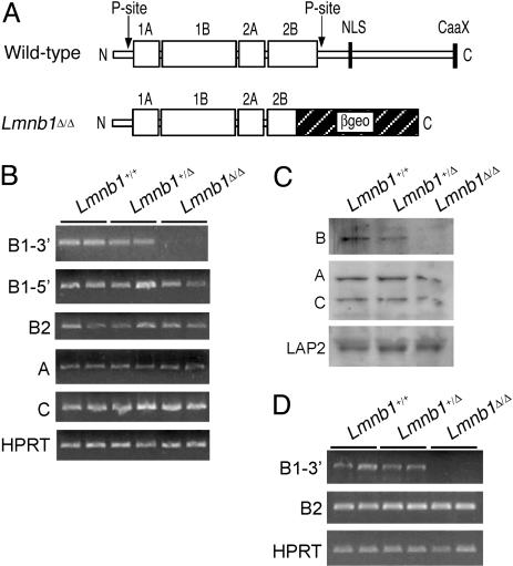
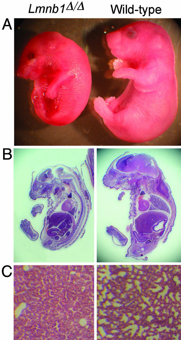
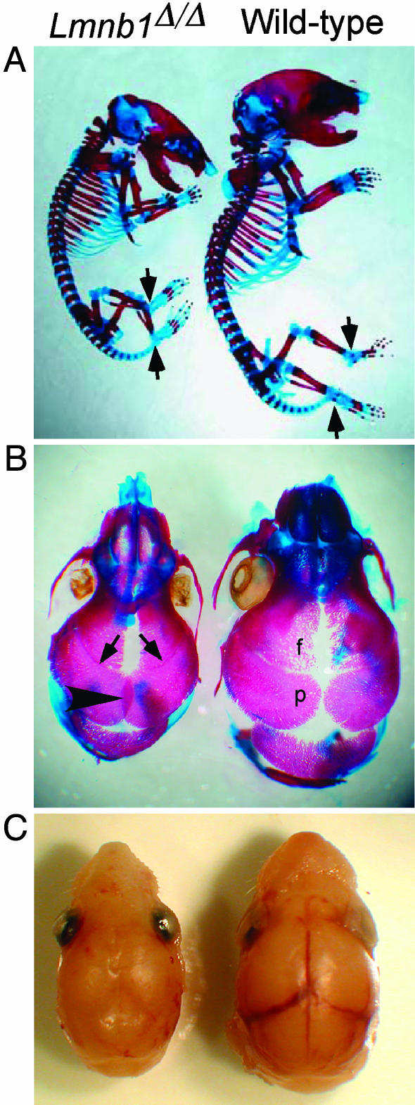
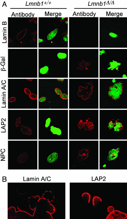
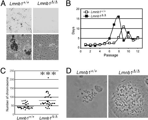
Lamins are key structural components of the nuclear lamina, an intermediate filament meshwork that lies beneath the inner nuclear membrane. Lamins play a role in nuclear architecture, DNA replication, and gene expression. Mutations affecting A-type lamins have been associated with a variety of human diseases, including muscular dystrophy, cardiomyopathy, lipodystrophy, and progeria, but mutations in B-type lamins have never been identified in humans or in experimental animals. To investigate the in vivo function of lamin B1, the major B-type lamin, we generated mice with an insertional mutation in Lmnb1. The mutation resulted in the synthesis of a mutant lamin B1 protein lacking several key functional domains, including a portion of the rod domain, the nuclear localization signal, and the CAAX motif (the carboxyl-terminal signal for farnesylation). Homozygous Lmnb1 mutant mice survived embryonic development but died at birth with defects in lung and bone. Fibroblasts from mutant embryos grew under standard cell-culture conditions but displayed grossly misshapen nuclei, impaired differentiation, increased polyploidy, and premature senescence. Thus, the lamin B1 mutant mice provide evidence for a broad and nonredundant function of lamin B1 in mammalian development. These mutant mice and cell lines derived from them will be useful models for studying the role of the nuclear lamina in various cellular processes.
Figures
Similar articles
-
Do lamin B1 and lamin B2 have redundant functions?Nucleus. 2014 Jul-Aug;5(4):287-92. doi: 10.4161/nucl.29615. Nucleus. 2014. PMID: 25482116 Free PMC article.
-
Disruption of lamin B1 and lamin B2 processing and localization by farnesyltransferase inhibitors.Nucleus. 2013 Mar-Apr;4(2):142-50. doi: 10.4161/nucl.24089. Epub 2013 Mar 1. Nucleus. 2013. PMID: 23475125 Free PMC article.
-
Nuclear envelope remodelling during human spermiogenesis involves somatic B-type lamins and a spermatid-specific B3 lamin isoform.Mol Hum Reprod. 2015 Mar;21(3):225-36. doi: 10.1093/molehr/gau111. Epub 2014 Dec 4. Mol Hum Reprod. 2015. PMID: 25477337
-
Laminopathies and the long strange trip from basic cell biology to therapy.J Clin Invest. 2009 Jul;119(7):1825-36. doi: 10.1172/JCI37679. Epub 2009 Jul 1. J Clin Invest. 2009. PMID: 19587457 Free PMC article. Review.
-
The role of lamin B1 for the maintenance of nuclear structure and function.Nucleus. 2015;6(1):8-14. doi: 10.1080/19491034.2014.1003510. Nucleus. 2015. PMID: 25602590 Free PMC article. Review.
Cited by
-
The nuclear lamina regulates germline stem cell niche organization via modulation of EGFR signaling.Cell Stem Cell. 2013 Jul 3;13(1):73-86. doi: 10.1016/j.stem.2013.05.003. Cell Stem Cell. 2013. PMID: 23827710 Free PMC article.
-
Age-dependent changes in nuclear-cytoplasmic signaling in skeletal muscle.Exp Gerontol. 2021 Jul 15;150:111338. doi: 10.1016/j.exger.2021.111338. Epub 2021 Apr 17. Exp Gerontol. 2021. PMID: 33862137 Free PMC article.
-
Defects in lamin B1 expression or processing affect interphase chromosome position and gene expression.J Cell Biol. 2007 Feb 26;176(5):593-603. doi: 10.1083/jcb.200607054. Epub 2007 Feb 20. J Cell Biol. 2007. PMID: 17312019 Free PMC article.
-
A Nuclear Belt Fastens on Neural Cell Fate.Cells. 2022 May 27;11(11):1761. doi: 10.3390/cells11111761. Cells. 2022. PMID: 35681456 Free PMC article. Review.
-
Nuclear envelope and lamin B2 function in the central nervous system.Proc Natl Acad Sci U S A. 2010 Apr 6;107(14):6121-2. doi: 10.1073/pnas.1000863107. Epub 2010 Apr 1. Proc Natl Acad Sci U S A. 2010. PMID: 20360557 Free PMC article. No abstract available.
References
-
- Cohen, M., Lee, K. K., Wilson, K. L. & Gruenbaum, Y. (2001) Trends Biochem. Sci. 26, 41–47. - PubMed
-
- Wilson, K. L., Zastrow, M. S. & Lee, K. K. (2001) Cell 104, 647–650. - PubMed
-
- Goldman, R. D., Gruenbaum, Y., Moir, R. D., Shumaker, D. K. & Spann, T. P. (2002) Genes Dev. 16, 533–547. - PubMed
-
- Burke, B. & Stewart, C. L. (2002) Nat. Rev. Mol. Cell. Biol. 3, 575–585. - PubMed
-
- Hutchison, C. J. (2002) Nat. Rev. Mol. Cell. Biol. 3, 848–858. - PubMed
Publication types
MeSH terms
Substances
Grants and funding
LinkOut - more resources
Full Text Sources
Other Literature Sources
Molecular Biology Databases
Research Materials
