. 2004 Jul;114(1):49-56.
doi: 10.1172/JCI21655.

Neutrophil protein kinase Cdelta as a mediator of stroke-reperfusion injury

Affiliations

Neutrophil protein kinase Cdelta as a mediator of stroke-reperfusion injury

Wen-Hai Chou et al. J Clin Invest. 2004 Jul.

Abstract

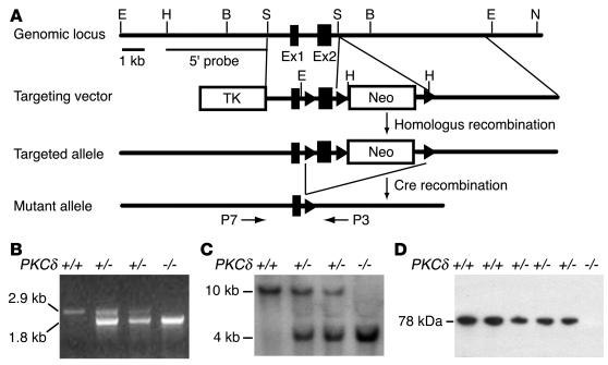
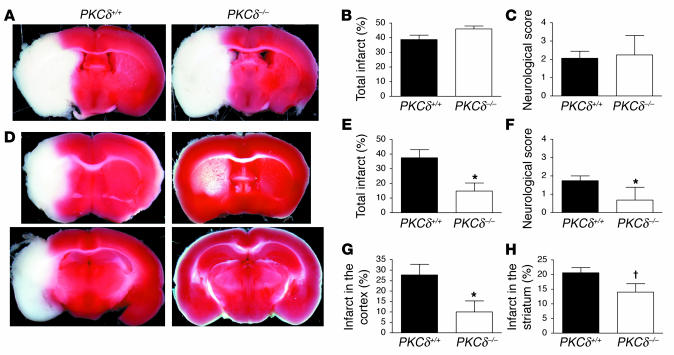
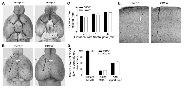
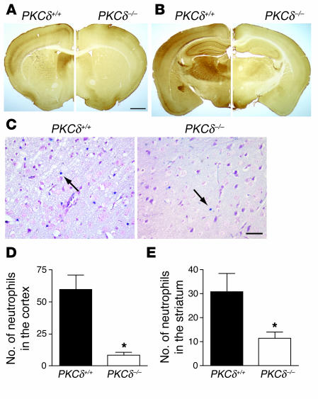
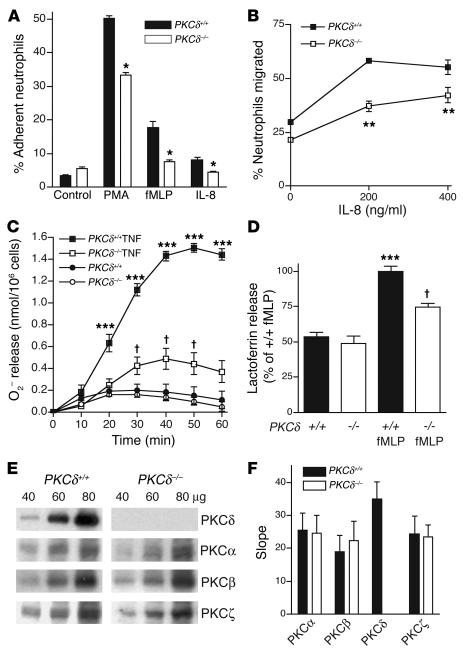
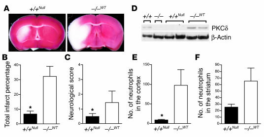
Thrombolysis is widely used to intervene in acute ischemic stroke, but reestablishment of circulation may paradoxically initiate a reperfusion injury. Here we describe studies with mice lacking protein kinase Cdelta (PKCdelta) showing that absence of this enzyme markedly reduces reperfusion injury following transient ischemia. This was associated with reduced infiltration of peripheral blood neutrophils into infarcted tissue and with impaired neutrophil adhesion, migration, respiratory burst, and degranulation in vitro. Total body irradiation followed by transplantation with bone marrow from PKCdelta-null mice donors reduced infarct size and improved neurological outcome in WT mice, whereas marrow transplantation from WT donors increased infarction and worsened neurological scores in PKCdelta-null mice. These results indicate an important role for neutrophil PKCdelta in reperfusion injury and strongly suggest that PKCdelta inhibitors could prove useful in the treatment of stroke.


Figures

Figure 1
Figure 1
Generation of PKCδ-null mice. (A) Organization of the mouse PKCδ gene, the targeting construct, and the allele resulting from homologous recombination. Boxes represent exon 1 (123 bp) and exon 2 (200 bp). Arrowheads represent loxP sequences. Only relevant restriction enzyme sites are indicated. E, EcoRI; H, HindIII; B, BamHI; S, SacI; N, NotI. (B) Verification of genotypes by PCR using the primers P7 and P3. The WT allele (+/+) generates a 2.9-kb and the mutant allele (−/−) a 1.8-kb band, as indicated. (C) Verification of genotypes by Southern blot analysis of genomic DNA digested with EcoRI from WT (+/+), heterozygous (+/ –), and homozygous mutants ( –/ –) using a 3.2-kb 5′-probe (HindIII-SacI fragment). (D) Verification of genotypes by Western blot analysis of whole brain lysates using a mAb against PKCδ. The migration of PKCδ immunoreactivity (78 kDa) is indicated.
Figure 2
Figure 2
Infarct size is reduced in PKCδ-null mice after transient, but not permanent, MCAO. (A) Shown are representative images of TTC-stained brain slices after 20 hours of permanent MCAO from PKCδ+/+ and PKCδ –/ – mice. Viable tissue is stained red, whereas the ischemic area remains unstained (white). (B) Infarct size measured as a percentage of the area of the nonischemic hemisphere after 20 hours of permanent MCAO from PKCδ+/+ (n = 8) and PKCδ –/ – mice (n = 8). (C) Neurological deficit scores after 20 hours of permanent MCAO from PKCδ+/+ (n = 8) and PKCδ –/ – mice (n = 8). (D) Representative images of TTC-stained brain slices after 1 hour of MCAO and 24 hours of reperfusion from PKCδ+/+ and PKCδ –/ – mice. (E) Total infarct size after 1 hour of MCAO and 24 hours of reperfusion from PKCδ+/+ (n = 10) and PKCδ –/ – (n = 9) mice. (F) Neurological deficit scores after 1 hour of MCAO and 24 hours of reperfusion from PKCδ+/+ (n = 8) and PKCδ –/ – mice (n = 8). (G and H) Infarct size in the cerebral cortex (G) and striatum (H) after 1 hour of MCAO and 24 hours of reperfusion from PKCδ+/+ (n = 10) and PKCδ –/ – (n = 9) mice. *P < 0.05 compared with WT littermates (two-tailed, unpaired t test). P < 0.05 compared with WT littermates (one-tailed, unpaired t test).
Figure 3
Figure 3
Cerebrovascular anatomy and cerebral blood flow. (A and B) Shown are representative images of the ventral (A) and dorsal (B) surfaces of brains from PKCδ+/+ and PKCδ –/ – mice perfused with black ink. The points of anastomoses between the MCA and the ACA are circled and connected by the line of anastomoses to define the respective vascular territories. (C) Distances from the line of anastomoses to the midline in PKCδ+/+ (n = 3) and PKCδ –/ – (n = 3) mice were measured at coronal planes 2, 4, and 6 mm from the frontal pole. (D) Regional cerebral blood flow before and during 1 hour of MCAO and during the first hour of reperfusion in PKCδ+/+ (n = 3) and PKCδ –/ – (n = 3) mice. Relative cerebral blood flow was expressed as the percentage of the Doppler signal intensity of the ischemic compared with the contralateral hemisphere. (E) The microvasculature in the cortex of PKCδ+/+ and PKCδ –/ – mice revealed by NADPH diaphorase histochemistry. The scale bar corresponds to 250 & μ;m.
Figure 4
Figure 4
Expression pattern of PKCδ in the brain and extravascular neutrophils after transient MCA occlusion. (A and B) Immunocytochemical localizations of PKCδ in the mouse brain are shown in coronal sections at bregma 0.38 mm (A) and a more posterior section at bregma –1.58 mm (B) for PKCδ+/+ (left) and PKCδ –/ – (right) mice. (C) Representative sections from PKCδ+/+ and PKCδ –/ – mice showing reduced neutrophil accumulation within infarcted tissue in the cortex of PKCδ –/ – mice after 1 hour of MCAO and 24 hours of reperfusion. Blue infiltrated neutrophils were identified in the ischemic cortex by staining for esterase activity with dichloroacetate (arrows). (D and E) Number of extravascular neutrophils in the ischemic cortex and striatum of PKCδ+/+ (n = 7) and PKCδ –/ – mice (n = 6) after transient MCAO. No esterase staining was seen in sections from nonischemic animals (data not shown). The scale bars in A and C correspond to 1 mm and 25 & μ;m, respectively. *P < 0.05 compared with WT littermates.
Figure 5
Figure 5
Decreased neutrophil function in PKCδ-null mice. (A) Neutrophil adhesion stimulated by PMA (100 nM), fMLP peptide (1 & μ;M), or IL-8 (200 ng/ml). *P < 0.05 compared with WT littermates (two-tailed, unpaired t tests). (B) IL-8–stimulated neutrophil migration differed by genotype [F(1, 6) = 54.8; P = 0.0003] and treatment [F(2, 6) = 63.08; P < 0.0001] without significant interaction between these factors [F(2, 6) = 3.9; NS]. **P < 0.05 compared with PKCδ+/+ at same dose (Bonferroni test). (C) TNF-α–stimulated (20 ng/ml) superoxide anion production showed main effects of treatment [F(1, 224) = 236; P < 0.0001], genotype [F(1, 224) = 506; P < 0.0001], and time [F(6, 224) = 64.6; P < 0.0001] with an interaction between these factors [F(6, 224) = 14.7; P < 0.0001]. P < 0.05 compared with unstimulated PKCδ –/ – neutrophils, and ***P < 0.05 compared with unstimulated PKCδ+/+ and stimulated PKCδ –/ – neutrophils at the same time (Newman Keuls test). (D) fMLP-stimulated (10 & μ;M) lactoferrin release differed by genotype [F(1, 12) = 15.2; P = 0.0021] and treatment [F(1, 12) = 89.01; P < 0.0001] with an interaction between these factors [F(1, 12) = 7,24; P = 0.0197]. P < 0.05 compared with unstimulated PKCδ –/ – neutrophils, and ***P < 0.05 compared with unstimulated PKCδ+/+ and stimulated PKCδ –/ – neutrophils (Bonferroni tests). (E) PKC isozyme immunoreactivity in neutrophils. (F) PKC isozyme immunoreactivity determined by regression analysis of protein concentrations and corresponding OD values on Western blots (n = 3). Assays of neutrophil function (AD) were performed in triplicate or quadruplicate from at least three animals of each genotype.
Figure 6
Figure 6
Bone marrow transplantation reverses phenotypes in PKCδ WT and null mice. (AC) Representative images of TTC-stained brain slices (A), infarct sizes (B), and neurological scores (C) after transient MCAO of irradiated WT mice transplanted with PKCδ-null bone marrow [(+/+NULL), n = 7] and irradiated null mice transplanted with WT marrow [( –/ –WT), n = 8]. (D) PKCδ immunoreactivity in bone marrow cells from transplanted mice to confirm the success of bone marrow transplantation. PKCδ immunoreactivity was detected in the marrow of WT (+/+) mice and irradiated null mice transplanted with WT marrow ( –/ –WT), but not in PKCδ-null mice ( –/ –) nor in irradiated WT mice transplanted with PKCδ-null bone marrow (+/+NULL). Shown are samples from one WT, one PKCδ-null, two +/+NULL, and two –/ –WT mice. Samples were normalized to β-actin immunoreactivity. (E and F) Number of extravascular neutrophils in the ischemic cortex (E) and striatum (F) of irradiated WT mice transplanted with PKCδ-null bone marrow [(+/+NULL), n = 6] and irradiated null mice transplanted with WT marrow [( –/ –WT), n = 6] after 1 hour of MCAO and 24 hours of reperfusion. *P < 0.05 compared with WT littermates.

Similar articles

Cited by

References

    1. Stapf C, Mohr JP. Ischemic stroke therapy. Annu. Rev. Med. 2002;53:453–475. - PubMed
    1. Becker KJ. Targeting the central nervous system inflammatory response in ischemic stroke. Curr. Opin. Neurol. 2001;14:349–353. - PubMed
    1. Emsley HC, Tyrrell PJ. Inflammation and infection in clinical stroke. J. Cereb. Blood Flow Metab. 2002;22:1399–1419. - PubMed
    1. Jean WC, Spellman SR, Nussbaum ES, Low WC. Reperfusion injury after focal cerebral ischemia: the role of inflammation and the therapeutic horizon. Neurosurgery. 1998;43:1382–1396. - PubMed
    1. Cavanagh SP, Gough MJ, Homer-Vanniasinkam S. The role of the neutrophil in ischaemia-reperfusion injury: potential therapeutic interventions. Cardiovasc. Res. 1998;6:112–118. - PubMed

Publication types