Comparative Study
. 2004 Sep 1;559(Pt 2):543-54.
doi: 10.1113/jphysiol.2004.065219. Epub 2004 Jul 2.

Changes in AMPA receptor currents following LTP induction on rat CA1 pyramidal neurones

Affiliations
Comparative Study

Changes in AMPA receptor currents following LTP induction on rat CA1 pyramidal neurones

Bertalan K Andrásfalvy et al. J Physiol. .

Abstract

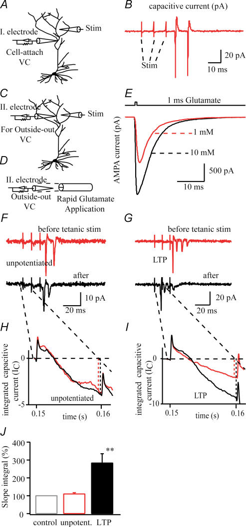
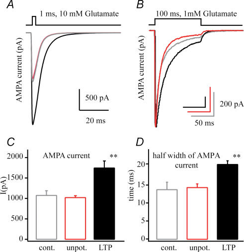
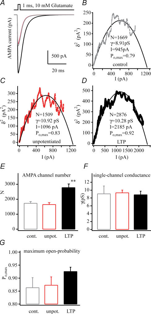
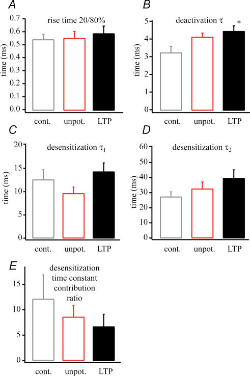
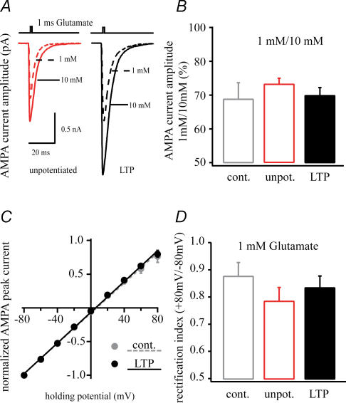
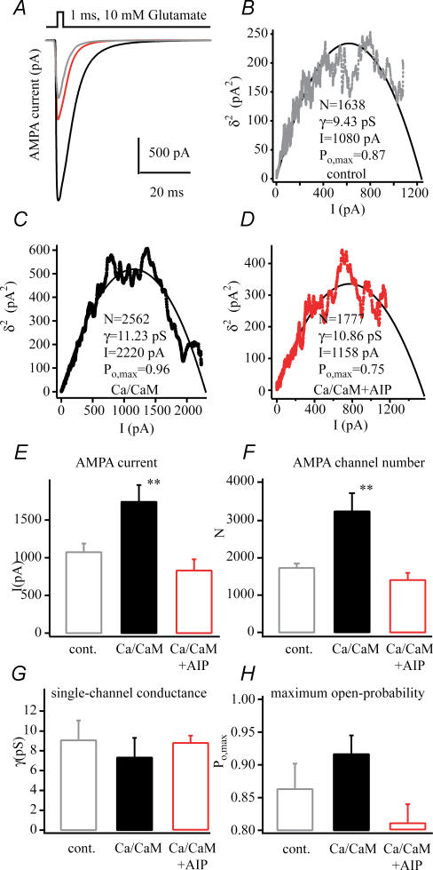
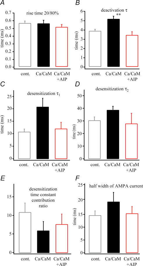
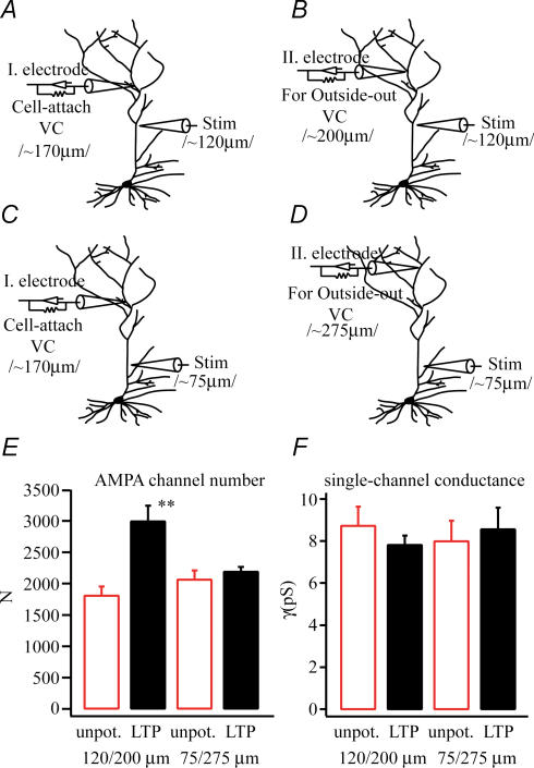
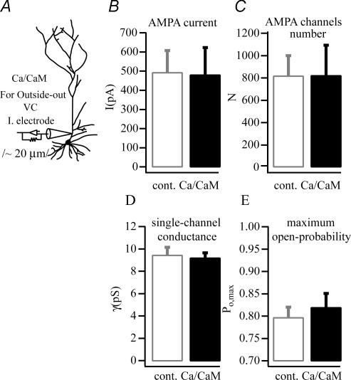
In the CA1 region of the hippocampus, LTP is thought to be initiated by a transient activation of NMDA receptors and is expressed as a persistent increase in synaptic transmission through AMPA receptors. To investigate the postsynaptic modifications of AMPA receptors involved in this enhanced synaptic transmission, the channel density and single-channel properties of extrasynaptic AMPA receptors located in synaptically active dendritic regions were examined following the induction of LTP. Following tetanic stimulation an outside-out patch was excised from the apical dendrite near the point of stimulation and saturating concentrations of glutamate were rapidly applied to the patch. AMPA current amplitude and duration were increased significantly in patches pulled from dendrites that expressed LTP. Non-stationary fluctuation analysis of AMPA currents indicated that AMPA channel number was nearly twofold larger than in controls, while single channel conductance and maximum open-probability were unchanged. Furthermore, while subtle changes in AMPA channel kinetics could also be observed, we did not find any evidence that receptor affinity or rectification properties were altered by LTP induction. Very similar results were found when CaMK-II activity was increased through the intracellular application of Ca/CaM. Together, we interpret our data to indicate that the stimuli used here produce an increased delivery of AMPA receptors to synaptically active regions of the apical dendrite without inducing any significant changes in their basic biophysical properties and that such delivery is a key element in this form of synaptic plasticity.


Figures

Figure 1
Figure 1
A, diagram of a dendritic cell-attached patch recording from ∼150 μm distal from the soma. B, representative trace of capacitive currents evoked by three test stimuli using stimulating electrode ∼50 μm far from the recording electrode. Test stimuli were given every 20 s, for 10 min before and 20 min after 100 Hz tetanic stimulation to measure the synaptic efficacy (FJ). C, diagram of second recording electrode ∼20 μm from stimulating electrode and ∼200 μm from soma to excise an outside-out patch for fast glutamate application (D). E, representative traces of AMPA receptor-mediated glutamate current (Vh = –80 mV) induced by 1 ms, 1 mm or 10 mm glutamate. Glutamate at 1 mm was used to measure kinetic properties (Fig. 4) and current–voltage relationship of AMPA receptors (Fig. 5). F and G, cell-attached patch recordings from dendrites before and after tetanic stimulation without (G) and with (F) NMDA receptor blockers evoked by three test stimuli. H and I, the integration of these capacitive currents (IC; caused by evoked EPSPs) using the average of three traces to measure the synaptic efficacy change caused by tetanic stimulation. J, the change of synaptic efficacy measuring peak deflection after the first test stimulus in time before and after tetanic stimulation (see text, **P < 0.01).
Figure 2
Figure 2. AMPA current changes following LTP
A, representative average AMPA currents from patches pulled from a control (grey), an unpotentiated neurone (50 μm APV and 10 μm MK-801, red) and a potentiated neurone (100 Hz tetanus, black) showing the increase in current amplitude that selectively occurs following LTP induction. B, average AMPA currents evoked by 100 ms 1 mm glutamate application showing the increased current duration following potentiation. C, numerical representation of the AMPA current amplitude of patches excised from control (n = 7), unpotentiated (n = 12) and potentiated (n = 11) neurones. D, half-width of AMPA currents quantifying the significant increase of AMPA current duration followed by LTP (**P < 0.01).
Figure 3
Figure 3. AMPA channel biophysical properties following LTP
A, representative average AMPA currents from patches pulled from a control (grey), an unpotentiated neurone (50 μm APV and 10 μm MK-801, red), and a potentiated neurone (100 Hz tetanus, black). BD, plots of mean AMPA current versus variance that were used in non-stationary fluctuation analyses of the currents shown above. The number of channels in the patch (N), single channel conductance (γ), mean peak current amplitude (I) and maximum channel open probability (Po,max) calculated from the fit of the plots by a parabolic function are shown (see methods). Pooled data of AMPA channel numbers (E) of dendritic excised patches from control (n = 6), unpotentiated (n = 12) and potentiated cells (n = 11, **P < 0.01). F, single-channel conductance (γ) appears equal in every condition. G, the maximum open probability (Po,max) shows a slight but statistically insignificant increase in the potentiated cells. The results of non-stationary fluctuation analysis suggest the channel number changes instead of the channel properties during potentiation. All dendritic outside-out patches were excised ∼200 μm distal to the soma, 25–35 min after 100 Hz tetanic stimulation. Glutamate (1 ms, 10 mm) was used for maximal saturation of AMPA receptors for non-stationary fluctuation analysis.
Figure 4
Figure 4. AMPA current kinetic changes following LTP
The kinetic properties of the AMPA receptor currents. A, rise time. B, deactivation (measured at 1 ms, 1 mm glutamate application) time constant (τ). C, fast desensitization time constant (τ1). D, slow desensitization (measured at 100 ms, 1 mm glutamate application) time constant (τ2). E, the desensitization time constant contribution ratio. A slowing in kinetics can be seen during potentiation (n = 11, LTP versus control, *P < 0.05).
Figure 5
Figure 5. Potentiation does not change receptor affinity or channel rectification properties
A, representative traces of 1 mm and 10 mm glutamate- (1 ms pulse) induced AMPA receptor-mediated currents of excised dendritic patch from control (n = 6), unpotentiated (n = 12) and potentiated (n = 11) cells. The ratio of 1 mm and 10 mm glutamate current (B) are similar in all cases suggesting that the glutamate affinity of AMPA receptors did not change during potentiation. C, the current–voltage relationship of control (filled grey circle) and LTP (filled black circle). The unpotentiated cells were similar (data not shown). D, the rectification index (peak current at +80/–80 mV) does not show any differences between these states.
Figure 6
Figure 6. AMPA channel biophysical property changes following CaMK-II activation
A, representative average AMPA currents from patches pulled from a control (grey), a CaMK-II-activated neurone (20 min Ca2+/CaM, black) and AIP-blocked CaMK-II-activated neurone (20 min Ca2+/CaM + AIP, red). BD, plots of mean AMPA current versus variance that were used in non-stationary fluctuation analyses of the currents shown above. The number of channels in the patch (N), single channel conductance (γ), mean peak current amplitude (I) and maximum channel open probability (Po,max) calculated from the fit of the plots by a parabolic function are shown (see methods). E and F, pooled data of increased AMPA currents (E) and AMPA channel numbers (F) of dendritic excised patches from control (n = 7), CaMK-II (n = 10), CaMK-II + AIP (n = 6). G, single-channel conductance (γ) appears equal in every condition. H, the maximum open probability (Po,max) shows an insignificant increase in the CaMK-II-activated cells. The results of non-stationary fluctuation analysis suggest the channel number changes instead of the channel properties during potentiation. All dendritic outside-out patches were excised ∼200 μm distal to the soma, 20–30 min Ca2+/CaM intracellular application. Glutamate (1 ms, 10 mm) was used for maximal saturation of AMPA receptors for non-stationary fluctuation analysis (**P < 0.01).
Figure 7
Figure 7. AMPA current kinetic changes following CaMKII activation
The kinetic properties of the AMPA receptor current. A, rise time. B, deactivation (measured at 1 ms, 1 mm glutamate application) time constant (τ). C, fast desensitization time constant (τ1). D, slow desensitization time constant (τ2). E, the desensitization (measured at 100 ms, 1 mm glutamate application) time constant contribution ratio of dendritic excised patches from control (n = 7), CaMK-II (n = 10), CaMK-II + AIP (n = 6). A slowing in kinetics can be seen during Ca/CaM treatment. AIP application with Ca/CaM shows the same values as in control. F, half-width of AMPA currents demonstrate the increase of AMPA current decay followed by CaMK-II activation (**P < 0.01).
Figure 8
Figure 8. Regionalization of AMPA receptor changes
A and B, the experimental configuration for pulling the outside-out patch near the site of the LTP induction (80–100 μm distal to the stimulation site). C and D, the experimental configuration for pulling patches at a site that was distant to the stimulation site (∼200 μm distant). Pooled data (LTP n = 4, unpotentiated n = 3) show that AMPA channel number (E) is increased when the patch was pulled near the site of stimulation but not when it was far away. There were no changes in single-channel conductance under any conditions.
Figure 9
Figure 9. Synaptic structures are required for the increase in AMPA channel numbers caused by CaMK-II activation
A, intracellular application of Ca/CaM (n = 7) at the proximal part of the apical dendrites (∼20 μm away from soma) did not have an impact on (control n = 9) any properties of excised AMPA receptors, BE), suggesting the localization dependence of phosphorylation governs receptor insertion or lateral transportation.
Figure 10
Figure 10. Model of AMPA receptor redistribution or transportation into the subsynaptic and synaptic area during LTP induction in adult hippocampal neurones
The picture shows a distal apical dentritic trunk and on the magnified insert the scale bar indicates the average tip aperture of our pipettes. A, the dashed circle indicates the putative outside-out patch membrane area containing extrasynaptic and sub- or juxtasynaptic AMPA receptors. Under resting conditions the GluR2/3 (red) AMPA receptors are continuously exo- and endocytosed directly into the PSD (green line). Most of the GluR1/2 AMPA receptors (black) are mobile in the extra- and subsynaptic area and continuously moving in and out of the PSD by lateral drift, but are not anchored there. B, when Ca2+ flows through NMDA ion channels (blue) during depolarization, many phosphorylation cascades (e.g. CaMK-II) are initiated, which activate transporting and anchoring proteins (AMPA-anchoring linkage). AMPA-anchoring linkages that are set together during phosphorylation move the mobile GluR1/2 receptors towards the PSD where they are then anchored. Lateral drift of more extrasynaptic receptors makes more receptors available in the sub- or juxtasynaptic region. The number of GluR1/2 receptors in this region mirrors the increase in synaptic AMPA receptors.

References

    1. Andrásfalvy BK, Magee JC. Distance-dependent increase in AMPA receptor number in the dendrites of adult hippocampal CA1 pyramidal neurons. J Neurosci. 2001;21:9151–9159. - PMC - PubMed
    1. Andrásfalvy BK, Smith MA, Borchardt T, Sprengel R, Magee JC. Impaired regulation of synaptic strength in hippocampal neurons from GluR1-deficient mice. J Physiol. 2003;552:35–45. - PMC - PubMed
    1. Bannister NJ, Larkman AU. Dendritic morphology of CA1 pyramidal neurons from the rat hippocampus. II. Spine distribution. J Comp Neurol. 1995;360:161–171. - PubMed
    1. Benke TA, Lüthi A, Isaac JT, Collingridge GL. Modulation of AMPA receptor unitary conductance by synaptic activity. Nature. 1998;393:793–797. - PubMed
    1. Bliss TV, Collingridge GL. A synaptic model of memory: long-term potentiation in the hippocampus. Nature. 1993;361:31–39. - PubMed

Publication types

MeSH terms

Substances