Comparative Study
. 2004 Sep 1;559(Pt 2):651-62.
doi: 10.1113/jphysiol.2004.064816. Epub 2004 Jul 2.

Serotonin and cholecystokinin synergistically stimulate rat vagal primary afferent neurones

Affiliations
Comparative Study

Serotonin and cholecystokinin synergistically stimulate rat vagal primary afferent neurones

Y Li et al. J Physiol. .

Retraction in

Abstract

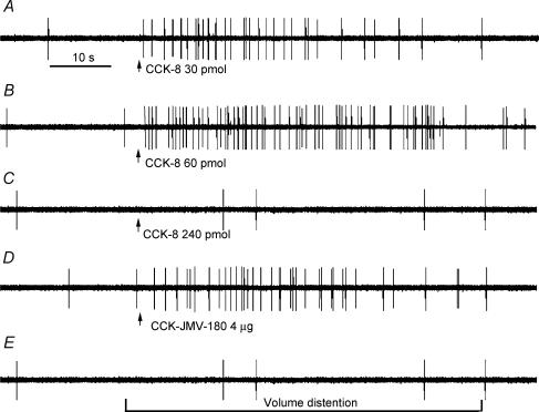
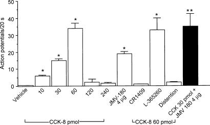
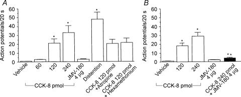
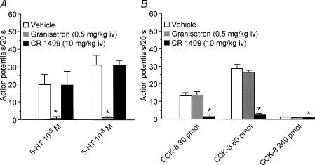
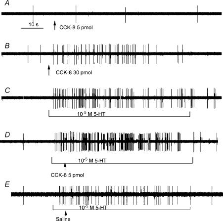
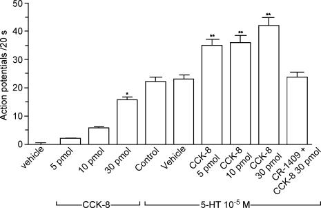
Recent studies indicate that cholecystokinin (CCK) and serotonin (5-hydroxytryptamine, 5-HT) act via vagal afferent fibres to mediate gastrointestinal functions. In the present study, we characterized the interaction between CCK and 5-HT in the vagal primary afferent neurones. Single neuronal discharges of vagal primary afferent neurones innervating the duodenum were recorded from rat nodose ganglia. Two groups of nodose ganglia neurones were identified: group A neurones responded to intra-arterial injection of low doses of cholecystokinin octapeptide (CCK-8; 10-60 pmol); group B neurones responded only to high doses of CCK-8 (120-240 pmol), and were also activated by duodenal distention. CCK-JMV-180, which acts as an agonist in high-affinity states and as an antagonist in low-affinity states, dose dependently stimulated group A neurones, but inhibited the effect of the high doses of CCK-8 on group B neurones. Duodenal perfusion of 5-HT evoked dose-dependent increases in nodose neuronal discharges. Some neurones that responded to 5-HT showed no response to either high or low doses of CCK-8. A separate group of nodose neurones that possessed high-affinity CCK type A (CCK-A) receptors also responded to luminal infusion of 5-HT. Further, a subthreshold dose of CCK-8 (i.e. 5 pmol) produced no measurable electrophysiological effects but it augmented the neuronal responses to 5-HT. This potentiation effect of CCK-8 was eliminated by CR 1409. From these results we concluded that the vagal nodose ganglion contains neurones that may possess only high- or low-affinity CCK-A receptors or 5-HT3 receptors. Some neurones that express high-affinity CCK-A receptors also express 5-HT3 receptors. Pre-exposure to luminal 5-HT may augment the subsequent response to a subthreshold dose of CCK.


Figures

Figure 1
Figure 1. Response of a nodose ganglia neurone to intra-superior pancreaticoduodenal artery infusions of CCK-8 and CCK-JMV-180 and to intestinal volume distention
Intra-arterial infusion of CCK-8 at doses of 30 pmol (A) and 60 pmol (B) produced a dose-dependent increase in nodose neuronal discharge frequency. C, administration of CCK-8 at 240 pmol evoked only a slight increase in nodose neuronal firing. D, administration of CCK-JMV-180 produced a marked increase in vagal afferent discharge in the same neurone as in A, which indicates the presence high-affinity CCK-A receptors on a subgroup of vagal primary afferent neurones. However, this same neurone failed to respond to intestinal distention (E).
Figure 2
Figure 2. Discharges of group A nodose ganglia neurones in response to intra-arterial injections of CCK-8 and CCK-JMV-180 and to intestinal volume distention
Of 141 neurones activated by electrical vagal stimulation, 24 neurones responded to intra-arterial injection of CCK-8 and CCK-JMV-180. Of these 24 neurones, two groups of neurones were identified. The discharge of group A neurones, which responded to low doses of CCK-8, were used for the analysis shown in this figure. Administration of CCK-8 at doses of 10, 30 and 60 pmol dose-dependently increased neuronal discharges. This group of neurones did not respond to high doses of CCK-8 (i.e. 120 and 240 pmol), and they failed to respond to intestinal volume distention. Twenty of 24 group A neurones were activated by intra-arterial injection of CCK-JMV-180. Administration of the CCK-A receptor antagonist CR 1409, but not the CCK-B receptor antagonist L-365,260, abolished nodose neuronal responses evoked by CCK-8 and CCK-JMV-180. Administration of CCK-JMV-180 in combination with CCK-8 (30 pmol) produced a further increase of neuronal firings compared with administration of CCK-8 alone. The filled bar represents the mean value of the neuronal discharges in response to CCK-8 (30 pmol) plus CCK-JMV-180. Values are means ± s.e.m. *P < 0.01 compared with vehicle; **P < 0.01 compared with CCK-8 at 30 pmol.
Figure 3
Figure 3. Discharges of group B nodose ganglia neurones in response to intra-arterial injections of CCK-8 and CCK-JMV-180 and to intestinal volume distention
A, discharges of the group B neurones that responded to high doses of CCK-8 (i.e. 120 and 240 pmol). Neither administration of CCK at low doses nor administration of CCK-JMV-180 stimulated this group of neurones. In contrast to group A neurones, intestinal distention markedly increased the neuronal discharge frequency. Administration of atropine or hexamethonium however, had no effect on neuronal discharge stimulated by CCK-8. B, in a separate study of the 12 neurones, five were activated by high doses of CCK-8. This group of neurones did not respond to CCK-JMV-180. Administration of CCK-JMV-180 produced an 80% inhibition of neuronal responses stimulated by the 240 pmol dose of CCK-8. The filled bar represents the mean values of the neuronal discharges in response to CCK-8 (240 pmol) plus CCK-JMV-180. *P < 0.01 compared with vehicle; **P < 0.01 compared with CCK-8 at 240 pmol.
Figure 4
Figure 4. Responses of nodose ganglia neurones activated by both intraluminal perfusion of 5-HT and intra-arterial injection of CCK-8
Of 32 neurones activated by electrical vagal stimulation, luminal perfusion of 5-HT (10−5 and 10−3 m) dose-dependently stimulated nodose neuronal firing in nine neurones A, administration of the 5-HT3 receptor antagonist granisetron but not the CCK-A receptor antagonist CR 1409, eliminated nodose neuronal responses evoked by luminal 5-HT stimulation. B, of the nine neurones that responded to luminal perfusion of 5-HT, five were activated by intra-arterial injection of low doses (i.e. 30 and 60 pmol) but not a high dose (240 pmol) of CCK-8. Administration of the CCK-A receptor antagonist CR 1409, but not the 5-HT3 receptor antagonist granisetron eliminated the neuronal responses evoked by CCK-8. Values are means ± s.e.m.
Figure 5
Figure 5. Effect of the interaction between CCK-8 and 5-HT on nodose neuronal firing
Intra-arterial infusion CCK-8 at a dose of 30 pmol (B), but not 5 pmol (A) increased nodose neuronal firing. Intraduodenal infusion of 10−5 m 5-HT stimulated the same neurone (C). D, administration of the subthreshold dose of CCK-8 (i.e. 5 pmol) enhanced the nodose neuronal response to 5-HT (10−5 m). E, administration of saline had no effect on the nodose neuronal response to 5-HT (10−5 m).
Figure 6
Figure 6. Discharge of nodose ganglia neurones in response to intra-arterial injection of CCK-8 and intraluminal perfusion of 5-HT, and the effect of the interaction between CCK-8 and 5-HT
Of the 40 neurones activated by electrical stimulation of the vagus nerve, a subthreshold dose of CCK (i.e. 5 pmol) produced no measurable electrophysiological effect. Eleven of 40 neurones were activated by intra-arterial injection of CCK at a dose of 30 pmol. Of the 11 neurones that responded to CCK-8, six were activated after luminal perfusion of 5-HT. Intra-arterial injection of a subthreshold dose of CCK (5 pmol) enhanced the neuronal responses to luminal 5-HT. Furthermore, injection of CCK (30 pmol) against a background luminal perfusion of 5-HT produced a further increase of neuronal firing. Administration of the CCK-A receptor antagonist CR 1409 eliminated the enhanced responses induced by CCK-8. *P < 0.01 compared with vehicle; **P < 0.01 compared with 5-HT- control.

Similar articles

Cited by

References

    1. Andrews PL, Davison JS. Activation of vagal afferent terminals by 5-HT is mediated by 5-HT3 receptors in the anaesthetized ferret. J Physiol. 1990;422:92P.
    1. Attoub S, Moizo L, Laignwau JP, Alchepo B, Lewin MJ, Bado A. YM022, a highly potent and selective CCKB antagonist inhibiting gastric acid secretion in the rat, the cat and isolated rabbit glands. Fundam Clin Pharmacol. 1998;12:256–262. - PubMed
    1. Blackshaw LA, Grundy D. Effects of 5-hydroxytryptamine on discharge of vagal mucosal afferent fibres from the upper gastrointestinal tract of the ferret. J Auton Nerv Syst. 1993;45:41–50. - PubMed
    1. Bueno L, Fioramonti J, Delvaux M, Frexinos J. Mediators and pharmacology of visceral sensitivity: from basic to clinical investigations. Gastroenterology. 1997;112:1714–1743. - PubMed
    1. Cox JE. Duodenal sucrose and glucose infusions enhance suppression by cholecystokinin of sham feeding. Am J Physiol. 1994;266:R466–R471. - PubMed

Publication types