The evolution of H5N1 influenza viruses in ducks in southern China
- PMID: 15235128
- PMCID: PMC478602
- DOI: 10.1073/pnas.0403212101
The evolution of H5N1 influenza viruses in ducks in southern China
Abstract
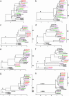
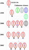
The pathogenicity of avian H5N1 influenza viruses to mammals has been evolving since the mid-1980s. Here, we demonstrate that H5N1 influenza viruses, isolated from apparently healthy domestic ducks in mainland China from 1999 through 2002, were becoming progressively more pathogenic for mammals, and we present a hypothesis explaining the mechanism of this evolutionary direction. Twenty-one viruses isolated from apparently healthy ducks in southern China from 1999 through 2002 were confirmed to be H5N1 subtype influenza A viruses. These isolates are antigenically similar to A/Goose/Guangdong/1/96 (H5N1) virus, which was the source of the 1997 Hong Kong "bird flu" hemagglutinin gene, and all are highly pathogenic in chickens. The viruses form four pathotypes on the basis of their replication and lethality in mice. There is a clear temporal pattern in the progressively increasing pathogenicity of these isolates in the mammalian model. Five of six H5N1 isolates tested replicated in inoculated ducks and were shed from trachea or cloaca, but none caused disease signs or death. Phylogenetic analysis of the full genome indicated that most of the viruses are reassortants containing the A/Goose/Guangdong/1/96-like hemagglutinin gene and the other genes from unknown Eurasian avian influenza viruses. This study is a characterization of the H5N1 avian influenza viruses recently circulating in ducks in mainland China. Our findings suggest that immediate action is needed to prevent the transmission of highly pathogenic avian influenza viruses from the apparently healthy ducks into chickens or mammalian hosts.
Figures


References
-
- Webby, R. & Webster, R. (2003) Science 302, 1519–1522. - PubMed
-
- Claas, E., Osterhaus, A., van Beek, R., De Jong, J., Rimmelzwaan, G., Senne, D., Krauss, S., Shortridge, K. & Webster, R. (1998) Lancet 351, 472–477. - PubMed
-
- Subbarao, K., Klimov, A., Katz, J., Regnery, H., Lim, W., Hall, H., Perdue, M., Swayne, D., Bender, C., Huang, J., et al. (1998) Science 279, 393–396. - PubMed
Publication types
MeSH terms
Associated data
- Actions
- Actions
- Actions
- Actions
- Actions
- Actions
- Actions
- Actions
- Actions
- Actions
- Actions
- Actions
- Actions
- Actions
- Actions
- Actions
- Actions
- Actions
- Actions
- Actions
- Actions
- Actions
- Actions
- Actions
- Actions
- Actions
- Actions
- Actions
- Actions
- Actions
- Actions
- Actions
- Actions
- Actions
- Actions
- Actions
- Actions
- Actions
- Actions
- Actions
- Actions
- Actions
- Actions
- Actions
- Actions
- Actions
- Actions
- Actions
- Actions
- Actions
- Actions
- Actions
- Actions
- Actions
- Actions
- Actions
- Actions
- Actions
- Actions
- Actions
- Actions
- Actions
- Actions
- Actions
- Actions
- Actions
- Actions
- Actions
- Actions
- Actions
- Actions
- Actions
- Actions
- Actions
- Actions
- Actions
- Actions
- Actions
- Actions
- Actions
- Actions
- Actions
- Actions
- Actions
- Actions
- Actions
- Actions
- Actions
- Actions
- Actions
- Actions
- Actions
- Actions
- Actions
- Actions
- Actions
- Actions
- Actions
- Actions
- Actions
- Actions
- Actions
- Actions
- Actions
- Actions
- Actions
- Actions
- Actions
- Actions
- Actions
- Actions
- Actions
- Actions
- Actions
- Actions
- Actions
- Actions
- Actions
- Actions
- Actions
- Actions
- Actions
- Actions
- Actions
- Actions
- Actions
- Actions
- Actions
- Actions
- Actions
- Actions
- Actions
- Actions
- Actions
- Actions
- Actions
- Actions
- Actions
- Actions
- Actions
- Actions
- Actions
- Actions
- Actions
- Actions
- Actions
- Actions
- Actions
- Actions
- Actions
- Actions
- Actions
- Actions
- Actions
- Actions
- Actions
- Actions
- Actions
- Actions
- Actions
- Actions
- Actions
- Actions
- Actions
- Actions
- Actions
- Actions
- Actions
Grants and funding
LinkOut - more resources
Full Text Sources
Other Literature Sources
Medical

