. 2004 Jul 13;101(28):10398-403.
doi: 10.1073/pnas.0403342101. Epub 2004 Jul 2.

Engagement of B7 on effector T cells by regulatory T cells prevents autoimmune disease

Affiliations

Engagement of B7 on effector T cells by regulatory T cells prevents autoimmune disease

Silke Paust et al. Proc Natl Acad Sci U S A. .

Abstract

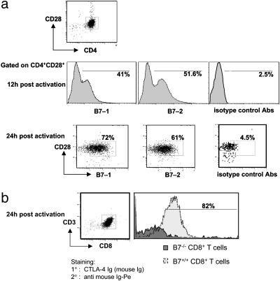
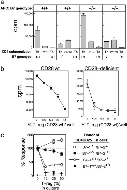
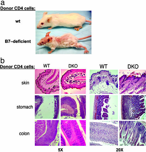
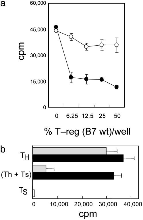
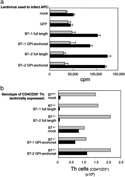
Although there is considerable evidence that a subpopulation of regulatory CD4(+)CD25+ T cells can suppress the response of autoreactive T cells, the underlying molecular mechanism is not understood. We find that transmission of a suppressive signal by CD4CD25+ regulatory cells requires engagement of the B7 molecule expressed on target T cells. The response of T cells from B7-deficient mice is resistant to suppression in vitro, and these cells provoke a lethal wasting disease in lymphopenic mice despite the presence of regulatory T cells. Susceptibility of B7-deficient cells to suppression is restored by lentiviral-based expression of full-length, but not truncated, B7 lacking a transmembrane/cytoplasmic domain. Because expression of these B7 truncation mutants restores CD28-dependent costimulatory activity, these findings that indicate B7-based transmission of suppressive activity suggest new approaches to modifying autoimmune responses.


Figures

Fig. 1.
Fig. 1.
Expression of B7-1 and B7-2 on Th (CD4CD25) or CD8+ T cells after activation. Th (CD4CD25) (a) or CD8+ T cells (b) from B7 wt or B7-deficient mice were stimulated with anti-CD3 on T cell-depleted irradiated spleen for the indicated time. Live cells were isolated and washed, and CD4 cells were stained with the indicated combinations of fluorescent-labeled Abs to CD4, CD28, and B7-1 or B7-2 or isotype control. CD8 cells were stained with CD3 and CTLA-4 Fc/nonlytic (mouse) followed by PE-labeled goat anti-mouse Ig. Isotype control Abs were biotinylated hamster IgG, group 1κ, and biotinylated rat IgG2b, κ, followed by streptavidin-PE (Pharmingen). Secondary Ab–streptavidin-PE staining without addition of primary Ab resulted in staining <2% (data not shown), and B7-deficient cells were used to demonstrate specificity of B7-1 and B7-2 staining.
Fig. 2.
Fig. 2.
Susceptibility of Th (CD4CD25) cells to suppression by T-reg (CD4CD25+). (a) Role of B7 expression on target Th cells. Purity of all cell isolations was ≥98%. Results shown are representative of three independent experiments. CD4CD25 cells (n = 2.5 × 10⁴, first and second panels) or CD4CD25 T cells (n = 5 × 10⁴, third and fourth panels) from B7 wt donors (first and third panels) or B7-deficient (second and fourth panels) and CD4CD25+ B7 wt T cells (n = 5 × 10⁴, all panels) were stimulated by themselves or mixed on 1 × 10⁵ irradiated B7 wt splenocytes (first and second panels) or 2 × 10⁵ irradiated B7-deficient splenocytes (third and fourth panels) with anti-CD3 mAb 2C11 (10 μg/ml). (b) Role of CD28 expression on target Th cells. Purity of all cell isolations was ≥98%. Results shown are representative of three independent experiments. CD4CD25 T cells (n = 5 × 10⁴) from CD28 wt donors (Left) or CD28-deficient donors (Right) and the indicated numbers of CD4CD25+ CD28 wt T cells were stimulated by themselves or admixed on 2 × 10⁵ irradiated B7 wt splenocytes with anti-CD3 mAb 2C11 (10 μg/ml). (c) Role of B7-1 versus B7-2 expression on target Th cells. Purity of all cell isolations was ≥98%. B7 wt (•), B7-deficient (□), B7-1-deficient/B7-2 wt (▪), and B7-1 wt/B7-2-deficient (○) CD4CD25 Th cells (n = 2.5 × 10⁴) were stimulated alone or in the presence of the indicated numbers of CD4CD25+ regulatory T cells on 1 × 10⁵ irradiated B7 wt spleen with soluble anti-CD3 2C11 (10 μg/ml). Data are shown as percent response: 100% corresponds to ≈80,000 cpm for B7-1/B7-1 wt Th and ranged from 30,000 to 50,000 for singly and doubly deficient Th cells. Proliferation was determined by measuring incorporation of [3H]thymidine (1 μCi per well) during the last 18 h of a 66-h culture by using scintillation counting.
Fig. 3.
Fig. 3.
Development of lethal wasting disease in RAG-2-deficient recipients of B7-deficient Th cells. (a) CD4+ cells (n = 106) purified from B7 wt or B7-deficient donor mice were transferred i.v. into syngeneic RAG-2-deficient recipients (n = 5) before being killed 30 days after transfer and preserved in Bouins' solution. Complete necropsies and disease scoring for gastritis and colitis (17, 18) were performed blind by the Harvard University Rodent Pathology Core. (b) Dermatitis is marked by inflammatory cells in the subdermal layer interspersed with hyperplastic endocrine cells and hyalinization of subepithelial space; gastritis by lymphocyte clusters in the mucosa, severe destruction of the mucosal architecture, disappearance of parietal and chief cells, and hyperplasia of mucous and endocrine cells; and colitis by accumulation of inflammatory cells between glands, mucin depletion from goblet cells, and marked thickening of the glands. DKO, double knock out (B7-1B7-2).
Fig. 4.
Fig. 4.
Absence of B7 from T cell targets, but not altered T cell development in B7-deficient mice, accounts for resistance to T-reg-mediated suppression. (a) BM chimera-derived B7 wt cells, but not B7-deficient Th cells, are susceptible to T-reg-mediated suppression. CD4CD25 cells (n = 2.5 × 10⁴) from BM chimeric mice reconstituted with B7-deficient BM (○) or B7 wt BM (•) and the indicated numbers of B7 wt CD4CD25+ T-reg were stimulated by themselves or admixed on 1 × 10⁵ irradiated B7 wt splenocytes with anti-CD3 mAb 2C11 (10 μg/ml). Purity of all cell isolations was ≥98%; results shown are representative of three independent experiments with a total of 10 mice per group. (b) Lentiviral expression of B7-1 reconstitutes susceptibility of B7-deficient Th cells to T-reg-mediated suppression. Empty vector pL6/V5 control transfectants (n = 2.5 × 10⁴, black bars) or B7-1-transfected CD4CD25 T cell targets (n = 2.5 × 10⁴, gray bars) were stimulated on 2 × 10⁵ B7 wt irradiated splenocytes with soluble anti-CD3 (10 μg/ml) in the presence or absence of 2.5 × 10⁴ B7 wt CD4CD25+ T-regs. Proliferation was determined by measuring incorporation of [3H]thymidine (1 μCi per well) during the last 18 h of a 66-h culture by using scintillation counting. Results are representative of two independent experiments.
Fig. 5.
Fig. 5.
Susceptibility of B7-deficient cells to suppression is restored by lentiviral-based expression of full-length, but not truncated, B7 lacking a transmembrane/cytoplasmic domain. (a) Examination of the costimulatory ability of cells infected with full-length or GPI-anchored B7-1 or B7-2. CD28 wt (black bars) or CD28–/– (gray bars) CD4CD25 T cells (n = 5 × 10⁴) were incubated with 1 × 10⁴ irradiated B7-deficient lentivirally infected dendritic cells in the presence of anti-CD3 (10 μg/ml). The ability of full-length or GPI-anchored B7-1 or B7-2 to provide costimulatory activity was determined from the CD28-dependent increase in proliferation. Proliferation was determined by measuring incorporation of [3H]thymidine (1 μCi per well) during the last 18 h of a 66-h culture by scintillation counting. Purity of all cell isolations was ≥98%. Results shown are representative of two independent experiments. (b) Lentiviral expression of B7-1 reconstitutes susceptibility of B7-deficient Th cells to T-reg-mediated suppression. Th (CD4CD25) mock- or lentivirally infected B7-deficient or B7 wt T cells (n = 106) were transferred intravenously into B7 wt RAG-2-deficient recipients (n = 4 per group) in the presence (black bars) or absence (gray bars) of 0.5 × 10⁶ CD4+CD25+ T cells. Recipients were killed 6 weeks after transfer, and the mandibular, cervical, axillary, inguinal, and mesenteric lymph nodes were then isolated. Total cell numbers were determined by trypan blue exclusion, and the percentage of CD4+ T cells was determined by FACS analysis. Transferred Th cells were ≥97% pure, and expression of lentivirally expressed B7 was ≥95% in all groups as confirmed by FACS analysis (Fig. 6). Data shown for each experimental group is the average of four pooled mice.

Similar articles

Cited by

References

    1. McHugh, R. S., Shevach, E. M. & Thornton, A. M. (2001) Microb. Infect. 3, 919–927. - PubMed
    1. Shimizu, J., Yamazaki, S., Takahashi, T., Ishida, Y. & Sakaguchi, S. (2002) Nat. Immunol. 3, 135–142. - PubMed
    1. Fontenot, J. D., Gavin, M. A. & Rudensky, A. Y. (2003) Nat. Immunol. 4, 330–336. - PubMed
    1. Hori, S., Nomura, T. & Sakaguchi, S. (2003) Science 299, 1057–1061. - PubMed
    1. Bystry, R. S., Aluvihare, V., Welch, K. A., Kallikourdis, M. & Betz, A. G. (2001) Nat. Immunol. 2, 1126–1132. - PubMed

MeSH terms