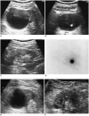
Percutaneous sclerotherapy of renal cysts with a beta-emitting radionuclide, holmium-166-chitosan complex
- PMID: 15235238
- PMCID: PMC2698141
- DOI: 10.3348/kjr.2004.5.2.128
Percutaneous sclerotherapy of renal cysts with a beta-emitting radionuclide, holmium-166-chitosan complex
Abstract
Objective: To evaluate the usefulness of a beta-emitting radionuclide (holmium-166-chitosan complex) as a sclerosing agent for the treatment of renal cysts.
Materials and methods: Using 10-30 mCi of holmium-166-chitosan complex, 20 renal cysts in 17 patients (14 male and 3 female patients, ranging in age from 47 to 82 years) were treated by percutaneous sclerotherapy under ultrasonographic guidance. The volume of the cysts before and after the sclerotherapy and the percentage change in volume were calculated in order to evaluate the response to therapy, which was classified as either complete regression (invisible), nearly complete regression (< 15 volume% of initial volume), partial regression (15-50 volume%) or no regression (> 50 volume%).
Results: The follow-up period ranged from 6 to 36 months (mean 28 months). Eighteen cysts (90%) regressed completely (n=11, 55%) or near-completely (n=7, 35%). Partial regression was obtained in one patient (5%) and there was no regression in one patient (5%). No significant complications were encountered.
Conclusion: The holmium-166-chitosan complex seems to be useful as a new painless sclerosing agent for the treatment of renal cysts with no significant complications.
Figures


Similar articles
-
Acetic acid as a sclerosing agent for renal cysts: comparison with ethanol in follow-up results.Cardiovasc Intervent Radiol. 2000 May-Jun;23(3):177-81. doi: 10.1007/s002700010039. Cardiovasc Intervent Radiol. 2000. PMID: 10821890 Clinical Trial.
-
MR evaluation of radiation synovectomy of the knee by means of intra-articular injection of holmium-166-chitosan complex in patients with rheumatoid arthritis: results at 4-month follow-up.Korean J Radiol. 2003 Jul-Sep;4(3):170-8. doi: 10.3348/kjr.2003.4.3.170. Korean J Radiol. 2003. PMID: 14530646 Free PMC article.
-
Efficacy of single-session percutaneous drainage and 50% acetic Acid sclerotherapy for treatment of simple renal cysts.Cardiovasc Intervent Radiol. 2007 Nov-Dec;30(6):1227-33. doi: 10.1007/s00270-007-9176-5. Epub 2007 Oct 4. Cardiovasc Intervent Radiol. 2007. PMID: 17914652
-
One-year results of single-session sclerotherapy with bleomycin in simple renal cysts.J Vasc Interv Radiol. 2012 Dec;23(12):1651-6. doi: 10.1016/j.jvir.2012.08.030. J Vasc Interv Radiol. 2012. PMID: 23177112 Clinical Trial.
-
[Percutaneous treatment of symptomatic renal cysts: effects of the combination of sclerotherapy with alcohol and fibrin glue (tissucol)].Radiol Med. 1993 Nov;86(5):657-61. Radiol Med. 1993. PMID: 8272552 Review. Italian.
Cited by
-
Alcohol sclerotherapy in the treatment of symptomatic simple renal cysts.Bosn J Basic Med Sci. 2008 Nov;8(4):337-40. doi: 10.17305/bjbms.2008.2893. Bosn J Basic Med Sci. 2008. PMID: 19125704 Free PMC article.
-
Radiologically guided percutaneous aspiration and sclerotherapy of symptomatic simple renal cysts: a systematic review of outcomes.Abdom Radiol (NY). 2021 Jun;46(6):2875-2890. doi: 10.1007/s00261-021-02953-9. Epub 2021 Feb 5. Abdom Radiol (NY). 2021. PMID: 33544165
-
The various therapeutic applications of the medical isotope holmium-166: a narrative review.EJNMMI Radiopharm Chem. 2019 Aug 5;4(1):19. doi: 10.1186/s41181-019-0066-3. EJNMMI Radiopharm Chem. 2019. PMID: 31659560 Free PMC article. Review.
-
Intracavitary radiation therapy for recurrent cystic brain tumors with holmium-166-chico : a pilot study.J Korean Neurosurg Soc. 2013 Sep;54(3):175-82. doi: 10.3340/jkns.2013.54.3.175. Epub 2013 Sep 30. J Korean Neurosurg Soc. 2013. PMID: 24278644 Free PMC article.
-
Comparison of acetic acid and ethanol sclerotherapy for simple renal cysts: clinical experience with 86 patients.Springerplus. 2016 Mar 9;5:299. doi: 10.1186/s40064-016-1971-5. eCollection 2016. Springerplus. 2016. PMID: 27066335 Free PMC article.
References
-
- Nascimento AB, Mitchell DG, Zhang X-M, Kamishima T, Parker L, Holland GA. Rapid MR imaging detection of renal cysts: age-based standards. Radiology. 2001;221:628–632. - PubMed
-
- Terada N, Ichioka K, Matsuta Y, Okubo K, Yoshimura K, Arai Y. The natural history of simple renal cysts. J Urol. 2002;167:21–23. - PubMed
-
- Pal DK, Kundu AK, Das S. Simple renal cysts: and observation. J Indian Med Assoc. 1997;95:555–558. - PubMed
-
- Rockson SG, Stone RA, Gunnells JC., Jr Solitary renal cyst with segmental ischemia and hypertension. J Urol. 1974;112:550–552. - PubMed
-
- Churchill D, Kimoff R, Pinsky M, et al. Solitary intrarenal cyst: correctable cause of hypertension. Urology. 1975;6:485–488. - PubMed
Publication types
MeSH terms
Substances
LinkOut - more resources
Full Text Sources
Medical

