Review
. 2004 Jul 5:2:46.
doi: 10.1186/1477-7827-2-46.

Comparative aspects of trophoblast development and placentation

Affiliations
Review

Comparative aspects of trophoblast development and placentation

Anthony M Carter et al. Reprod Biol Endocrinol. .

Abstract

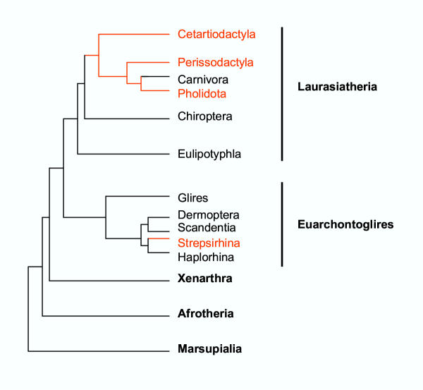
Based on the number of tissues separating maternal from fetal blood, placentas are classified as epitheliochorial, endotheliochorial or hemochorial. We review the occurrence of these placental types in the various orders of eutherian mammals within the framework of the four superorders identified by the techniques of molecular phylogenetics. The superorder Afrotheria diversified in ancient Africa and its living representatives include elephants, sea cows, hyraxes, aardvark, elephant shrews and tenrecs. Xenarthra, comprising armadillos, anteaters and sloths, diversified in South America. All placentas examined from members of these two oldest superorders are either endotheliochorial or hemochorial. The superorder Euarchontoglires includes two sister groups, Glires and Euarchonta. The former comprises rodents and lagomorphs, which typically have hemochorial placentas. The most primitive members of Euarchonta, the tree shrews, have endotheliochorial placentation. Flying lemurs and all higher primates have hemochorial placentas. However, the lemurs and lorises are exceptional among primates in having epitheliochorial placentation. Laurasiatheria, the last superorder to arise, includes several orders with epitheliochorial placentation. These comprise whales, camels, pigs, ruminants, horses and pangolins. In contrast, nearly all carnivores have endotheliochorial placentation, whilst bats have endotheliochorial or hemochorial placentas. Also included in Laurasiatheria are a number of insectivores that have many conserved morphological characters; none of these has epitheliochorial placentation. Consideration of placental type in relation to the findings of molecular phylogenetics suggests that the likely path of evolution in Afrotheria was from endotheliochorial to hemochorial placentation. This is also a likely scenario for Xenarthra and the bats. We argue that a definitive epitheliochorial placenta is a secondary specialization and that it evolved twice, once in the Laurasiatheria and once in the lemurs and lorises.


Figures

Figure 1
Figure 1
Eutherian fetal membranes. The embryo is enclosed in the amnion. Trophoblast (blue) and mesoderm (red) form the chorion. Trophoblast and yolk sac endoderm (yellow) together constitute a bilaminar omphalopleure. A choriovitelline or yolk sac placenta is then formed by interposition of mesoderm containing fetal blood vessels. Later the allantois expands into the exocoelom and the allantoic and chorionic mesoderm fuse to form a chorioallantoic placenta. The allantois will continue to expand into the exocoelom and eventually displace the yolk sac. The full sequence of events is seen most clearly in species with epitheliochorial placentation, such as horse and pig, although this cartoon is based on the rock hyrax (reference [82]).
Figure 2
Figure 2
Placentation in Afrotheria. (A) Endotheliochorial placenta of the African elephant, Loxodonta africana. The cellular trophoblast (CYTO TR) is deeply indented by the fetal capillary (FC), which is separated by <3 μm from the endothelium of the maternal capillary (ENDO). Reprinted from reference [23] with permission ©Elsevier 2003. (B). Hemochorial placenta of the lesser Madagascar hedgehog tenrec, Echinops telfairi. Maternal blood spaces (MBS) are lined by cellular trophoblast (CYTO TR) with microvilli (arrow). Reprinted from reference [33] with permission ©Elsevier 2003. (C) Fetal membranes in the rock hyrax Procavia capensis. The large allantoic sac is divided into four lobes by septa that carry four sets of allantoic vessels to the zonary placenta. The yolk sac is greatly reduced in late gestation. Reprinted from reference [83] with permission from the Carnegie Institution of Washington.
Figure 3
Figure 3
Placentation in Xenarthra. (A) Endotheliochorial placenta of the three-toed sloth, Bradypus tridactylus. The interhemal membrane comprises the enlarged maternal endothelial cells (ENDO), syncytial trophoblast (SYN TR) and the endothelium of the fetal capillary (FC). (B) Villous, hemomonochorial placenta of the nine-banded armadillo, Dasypus novemcinctus. The maternal blood space (MBS) is separated from the fetal capillary (FC) by syncytiotrophoblast (SYN TR).
Figure 4
Figure 4
Placentation in rodents (Glires). (A) Hemotrichorial placenta of the lemming, Lemmus lemmus. One layer of cellular trophoblast (T1) and two layers of syncytiotrophoblast (T2–T3) are interposed between the maternal blood space (MBS) and fetal capillaries (FC). (B) Hemomonochorial placenta of the golden-mantled ground squirrel, Spermophilus lateralis. The syncytiotrophoblast (SYN TR) has intrasyncytial bays (ISL). (C) Hemomonochorial placenta of a jumping mouse, Zapus sp. The interhemal membrane includes a single layer of giant trophoblast cells (CYTO TR). (D) Endotheliochorial placenta of a kangaroo rat, Dipodomys sp. The interhemal membrane comprises the endothelium of the maternal capillary (MC), cellular trophoblast (CYTO TR) with lipid inclusions and the endothelium of the fetal capillary (FC).
Figure 5
Figure 5
Placentation in primates (Euarchonta). (A) Epitheliochorial placenta of a bush baby, Galago crassicaudata. The interhemal membrane comprises maternal capillary (MC) endothelium, uterine epithelium (UT EP), cellular trophoblast (CYTO TR) and fetal capillary (FC) endothelium. (B) Endotheliochorial placenta of a tree shrew, Tupaia glis. The interhemal region contains maternal endothelium (MAT ENDO), syncytiotrophoblast (SYN TR) and the endothelium of the fetal capillary (FC). Reprinted from reference [52] with permission ©Springer-Verlag 1985. (C) The villous, hemomonochorial human placenta. The maternal blood space (MBS), or intervillous space, is lined by a thin layer of syncytiotrophoblast (SYN TR).
Figure 6
Figure 6
Placentation in bats and a ruminant. (A) Hemodichorial placenta of little brown bat, Myotis lucifugus (Vespertilionidae). There is an outer layer of syncytiotrophoblast (SYN TR) and an inner layer of cellular trophoblast (CYTO TR). Note the subsurface intrasyncytial spaces or bays (ISL) (B) Endotheliochorial placenta of funnel-eared bat, Natalus sp. (Natalidae) in late gestation. The interhemal region contains enlarged maternal endothelial cells (MAT ENDO) and cytotrophoblast (CYTO TR). (C) Hemomonochorial placenta of the Mexican free-tailed bat, Tadarida brasiliensis (Molossidae). The maternal blood spaces (MBS) are lined by cytotrophoblast (CYTO TR). (D) Epitheliochorial placenta of the sheep. The endometrial connective tissue has "decidual" cells whilst the fetal component comprises cellular trophoblast (T). Fusion of fetal and maternal cells gives rise to a syncytium (S). Therefore this type of placenta is sometimes referred to as synepitheliochorial. Reprinted from reference [8] with permission.
Figure 7
Figure 7
Cladogram based on nucleotide sequence analysis. The four superorders of eutherian mammals and the marsupial outgroup are shown in bold face. Epitheliochorial placentation, indicated in red, occurs in three orders of Laurasiatheria and one suborder of Euarchontoglires. These are the orders Cetartiodactyla (whales, pigs and hippopotami, camels, ruminants); Perissodactyla (horses, tapirs and rhinoceroses); Pholidota (pangolins or scaly anteaters); and the primate suborder Strepsirhina (lemurs and lorises).

Similar articles

Cited by

References

    1. Lillegraven JE. Polarities in mammalian evolution seen through homologs of the inner cell mass. J Mammal Evol. 2003;10:277–333. doi: 10.1023/B:JOMM.0000019774.11018.22. - DOI
    1. Freyer C, Zeller U, Renfree MB. The marsupial placenta: a phylogenetic analysis. J Exp Zoolog Part A Comp Exp Biol. 2003;299:59–77. - PubMed
    1. Blackburn DG, Johnson AR, Petzold JL. Histology of the extraembryonic membranes of an oviparous snake: towards a reconstruction of basal squamate patterns. J Exp Zoolog Part A Comp Exp Biol. 2003;299:48–58. - PubMed
    1. Cateni C, Paulesu L, Bigliardi E, Hamlett WC. The interleukin 1 (IL-1) system in the uteroplacental complex of a cartilaginous fish, the smoothhound shark, Mustelus canis. Reprod Biol Endocrinol. 2003;1:25. doi: 10.1186/1477-7827-1-25. - DOI - PMC - PubMed
    1. Flemming AF, Blackburn DG. Evolution of placental specializations in viviparous African and South American lizards. J Exp Zoolog Part A Comp Exp Biol. 2003;299:33–47. - PubMed

LinkOut - more resources