Comparative Study
. 2004 Aug;142(7):1171-81.
doi: 10.1038/sj.bjp.0705807. Epub 2004 Jul 5.

Calcitonin gene-related peptide (CGRP) modulates nociceptive trigeminovascular transmission in the cat

Affiliations
Comparative Study

Calcitonin gene-related peptide (CGRP) modulates nociceptive trigeminovascular transmission in the cat

Robin James Storer et al. Br J Pharmacol. 2004 Aug.

Abstract

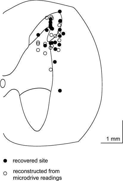
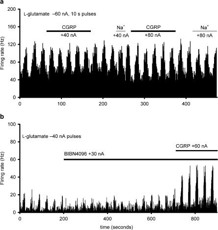
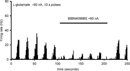
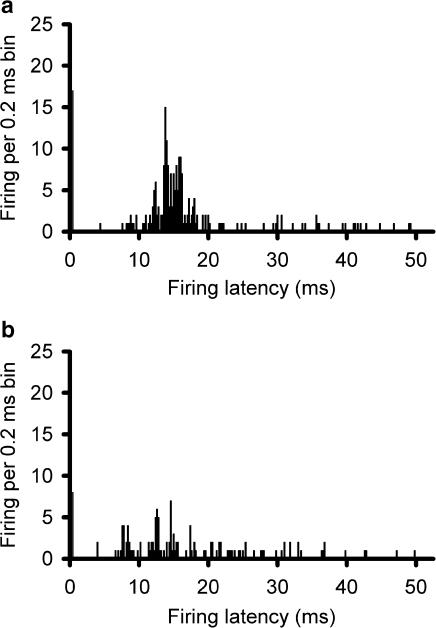
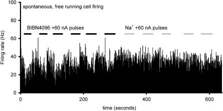
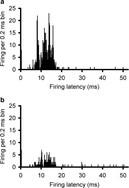
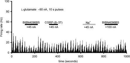
Calcitonin gene-related peptide (CGRP) is released into the cranial circulation of humans during acute migraine. To determine whether CGRP is involved in neurotransmission in craniovascular nociceptive pathways, we microiontophoresed onto neurons in the trigeminocervical complex and intravenously administered the CGRP receptor antagonists alpha-CGRP-(8-37) and BIBN4096BS. Cats were anaesthetised with alpha-chloralose, and using halothane during surgical preparation. A craniotomy and C1/C2 laminectomy allowed access to the superior sagittal sinus (SSS) and recording site. Recordings of activity in the trigeminocervical complex evoked by electrical stimulation of the SSS were made. Multibarrelled micropipettes incorporating a recording electrode were used for microiontophoresis of test substances. Cells recorded received wide dynamic range (WDR) or nociceptive specific (NS) input from cutaneous receptive fields on the face or forepaws. Cell firing was increased to 25-30 Hz by microiontophoresis of L-glutamate (n = 43 cells). Microiontophoresis of alpha-CGRP excited seven of 17 tested neurons. BIBN4096BS inhibited the majority of units (26 of 38 cells) activated by l-glutamate, demonstrating a non-presynaptic site of action for CGRP. alpha-CGRP-(8-37) inhibited a similar proportion of units (five of nine cells). Intravenous BIBN4096BS resulted in a dose-dependent inhibition of trigeminocervical SSS-evoked activity (ED50 31 microg kg(-1)). The maximal effect observed within 30 min of administration. The data suggest that there are non-presynaptic CGRP receptors in the trigeminocervical complex that can be inhibited by CGRP receptor blockade and that a CGRP receptor antagonist would be effective in the acute treatment of migraine and cluster headache.


Figures

Figure 1
Figure 1
Localisation of recording sites: A transverse section through the spinal cord at the level of C2 is represented by this schematic diagram. Microiontophoretically ejected Pontamine sky blue dye (6BX (C.I.24410)) was used to mark recording sites. Solid circles indicate sites where recording sites were identified histologically. The positions of unmarked recording sites, or sites where marks could not be recovered, were identified by reference to the position of dye marks at other recording sites and, or at the end of electrode tracts, and electrode tip coordinates, and are indicated by open circles. Although the recorded units are only mapped to one side of the cord in the schematic, they represent the data obtained from both the left-hand side and right-hand side of the spinal cord. The scale bar represents a distance of 1 mm in both directions.
Figure 2
Figure 2
Effect of α-CGRP and L-glutamate: L-glutamate (1.0 M, pH 8.0) was applied microiontophoretically (−60 nA) and increased the firing of trigeminocervical neurons linked to stimulation of the superior sagittal sinus. When the firing had reached a steady state over five epochs, α-CGRP (1 mM in 150 mM NaCl, pH 4.5) was co-microiontophoresed (solid line) with no added effect from sodium (+40 and +80 nA) in 10/17 units (panel a). In 7/17 units, trigeminal activity was increased by CGRP, albeit in the presence of BIBN4096BS (panel b). There was no significant effect of microiontophoresis of sodium. The histogram indicates the rate of firing seen in one second bins.
Figure 3
Figure 3
Effect of BIBN4096BS: Microiontophoresis of BIBN4096BS (20 mM, pH 5.7; +60 nA, solid bar) produces a robust reduction in L-glutamate-evoked (1.0 M, pH 8.0; 60 nA) firing in the trigeminocervical complex. The histogram indicates the rate of firing seen in one second bins.
Figure 4
Figure 4
Effect of BIBN4096BS microiontophoresis on SSS-evoked firing: Post-stimulus histograms showing that supramaximal electrical stimulation (50 × 250 μs) of the SSS via bipolar platinum hook electrodes recruits units in the trigeminocervical complex responding to the stimulus with a latency peak of 12 ms (panel a) that is inhibited immediately after microiontophoresis of BIBN4096BS (20 mM, pH 5.8; +60 nA for 300 s) (panel b). The apparent response within the first 0.2 ms is part of the stimulus artefact.
Figure 5
Figure 5
Effect of BIBN4096BS on spontaneous trigeminal neurons: Neurons firing spontaneously at a rate indicated in the histogram as firing per 1 s bin have a reduced firing frequency when BIBN4096BS is microiontophoretically ejected (20 mM, pH 5.7; +60 nA; black bars), while sodium (150 mM, pH 7.0) has no apparent effect at similar currents (grey bars).
Figure 6
Figure 6
Effect of intravenously administrated BIBN4096BS on SSS-evoked firing: Post-stimulus histograms showing that supramaximal electrical stimulation (50 × 250 μs) of the SSS via bipolar platinum hook electrodes recruits units in the trigeminocervical complex (panel a) that are substantially inhibited within 30 min of intravenous administration of BIBN4096BS (30 μg kg–1; panel b). The apparent response within the first 0.2 ms is part of the stimulus artefact.
Figure 7
Figure 7
Comparison of the effect of BIBN4096BS (20 mM, pH 5.8; +45 and +100 nA) and α-CGRP-(8 – 37) (1 mM in 160 mM NaCl, pH 4.5; +45 nA) in inhibiting the firing evoked by 10 s pulses of microiontophoretically applied L-glutamate (1.0 M, pH 8.0; −90 nA) onto a trigeminocervical complex unit linked to stimulation of the SSS, where sodium (150 mM; pH 7.0) has no apparent effect (+45 nA, grey bar). The firing rate indicated on the ordinate is indicated for 1 s bins by the rate histogram.

Comment in

Similar articles

Cited by

References

    1. AKERMAN S., WILLIAMSON D.J., KAUBE H., GOADSBY P.J. Nitric oxide synthase inhibitors can antagonise neurogenic and calcitonin gene-related peptide induced dilation of dural meningeal vessels. Br. J. Pharmacol. 2002;137:62–68. - PMC - PubMed
    1. ARMITAGE P, BERRY G. Statistical Methods in Medical Research 1994Oxford: Blackwell Science; 3rd edn
    1. BAHRA A., MATHARU M.S., BUCHEL C., FRACKOWIAK R.S.J., GOADSBY P.J. Brainstem activation specific to migraine headache. Lancet. 2001;357:1016–1017. - PubMed
    1. BEREITER D.A., BEREITER D.F., HATHAWAY C.B. The NMDA receptor antagonist MK-801 reduces Fos-like immunoreactivity in central trigeminal neurons and blocks select endocrine and autonomic responses to corneal stimulation in the rat. Pain. 1996;64:179–189. - PubMed
    1. BLOOM F.E. To spritz or not to spritz: the doubtful value of aimless iontophoresis. Life Sci. 1974;14:1819–1834. - PubMed

Publication types

MeSH terms

Substances