Comparative Study
. 2004 Jul 7:5:91.
doi: 10.1186/1471-2105-5-91.

SPOC: a widely distributed domain associated with cancer, apoptosis and transcription

Affiliations
Comparative Study

SPOC: a widely distributed domain associated with cancer, apoptosis and transcription

Luis Sánchez-Pulido et al. BMC Bioinformatics. .

Abstract

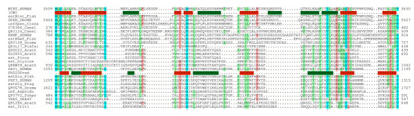
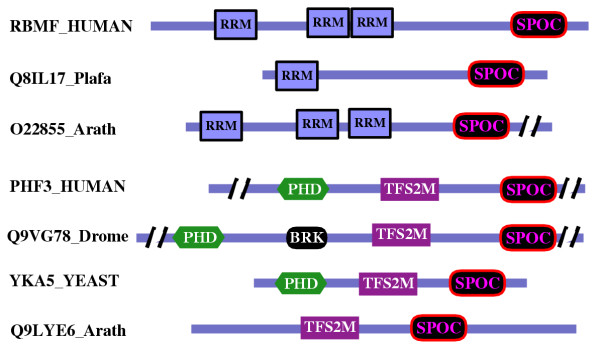
Background: The Split ends (Spen) family are large proteins characterised by N-terminal RNA recognition motifs (RRMs) and a conserved SPOC (Spen paralog and ortholog C-terminal) domain. The aim of this study is to characterize the family at the sequence level.

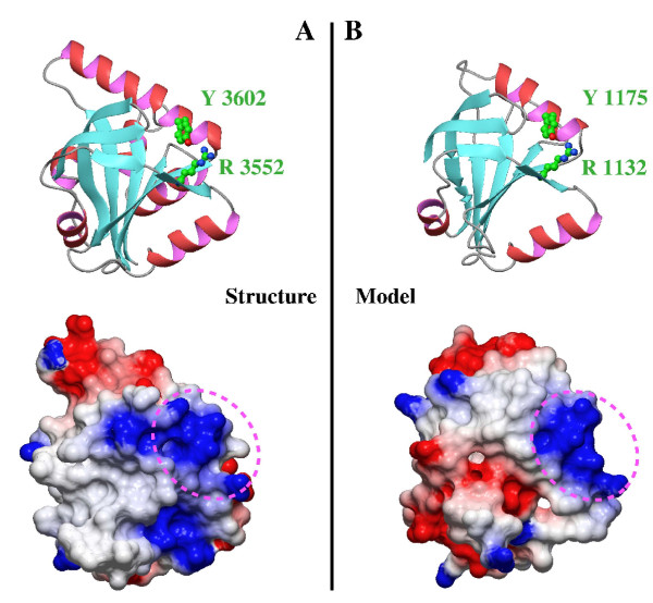
Results: We describe undetected members of the Spen family in other lineages (Plasmodium and Plants) and localise SPOC in a new domain context, in a family that is common to all eukaryotes using profile-based sequence searches and structural prediction methods.

Conclusions: The widely distributed DIO (Death inducer-obliterator) family is related to cancer and apoptosis and offers new clues about SPOC domain functionality.


Figures

Figure 1
Figure 1
Representative multiple alignment of the SPOC domain. The colouring scheme indicates average BLOSUM62 score (correlated to amino acid conservation) in each alignment column: cyan (greater than 3), light red (between 3 and 1.5) and light green (between 1.5 and 0.5). The limits of the domains are indicated by the residue positions on each side. The limits of proteins from partially sequenced genes whose full-length proteins are not available are not shown. X-Ray determined structure of the SPOC domain [9], pdb-code: 1OW1 is shown below the SHARP sequence (Swissprot-ID:MINT_HUMAN). PHD secondary structure prediction [29] for DIO family is shown below the DIO-1 human sequence (Swissprot-Id: DAT1_HUMAN), with E indicating a β strand (in red) and H an α helix (in green). The asterisks below the alignment marks the conserved pair arginine-tyrosine mentioned in the text. The sequences are named with their swissprot or sptrembl identifications, and also, if necessary, with their common species name: Human, Homo sapiens; Frog, Xenopus laevis; Drome, Drosophila melanogaster; Caeel, Caenorhabditis elegans; Arath,Arabidopsis thaliana; Ciona, Ciona intestinalis; Yeast, Saccharomyces cerevisiae; Fish, Brachydanio rerio; Plafal, Plasmodium falciparum; Aspnidu, Aspergillus nidulans; Pinus, Pinus taeda; Glycine, Glycine max; Schpo, Schizosaccharomyces pombe; Triti, Triticum aestivum. The "est" prefix identifies consensus sequences manually reconstructed by assembling highly similar expressed sequence tags from identical species (conceptual translations). The "unf" prefix identifies sequences obtained from Genome BLAST server at NCBI [24]. Complementary information is accessible at: .
Figure 2
Figure 2
Schematic representation of the domain architecture and common features in representative members of the SPOC domain contained families. The sequences are named with their swissprot or sptrembl identifications, and also, if necessary, with their common species name. The slashes represent inserts that are not shown. The proteins are drawn approximately to scale. The localization of the other domains PHD, TFIIS, BRK (BRM and KIS domain) and RRM is according to Pfam and SMART families databases [35,36].
Figure 3
Figure 3
Comparison of SPOC domains from SHARP and DIO proteins. (A) Ribbon representation and electrostatic surface potential map of structure of the SPOC domain of SHARP protein (PDB code: 1OW1). (B) Ribbon representation and electrostatic surface potential map of homology model of the SPOC domain of DIO protein. Blue indicates positively charged regions, whereas red shows negatively charged regions. All the molecules are in the same orientation. Dotted circles indicates the conserved basic cluster, where is located the conserved arginine-tyrosine pair.

References

    1. Chen F, Rebay I. split ends, a new component of the Drosophila EGF receptor pathway, regulates development of midline glial cells. Curr Biol. 2000;10:943–946. doi: 10.1016/S0960-9822(00)00625-4. - DOI - PubMed
    1. Kuang B, Wu SC, Shin Y, Luo L, Kolodziej P. split ends encodes large nuclear proteins that regulate neuronal cell fate and axon extension in the Drosophila embryo. Development. 2000;127:1517–1529. - PubMed
    1. Rebay I, Chen F, Hsiao F, Kolodziej PA, Kuang BH, Laverty T, Suh C, Voas M, Williams A, Rubin GM. A genetic screen for novel components of the Ras/Mitogen-activated protein kinase signaling pathway that interact with the yan gene of Drosophila identifies split ends, a new RNA recognition motif-containing protein. Genetics. 2000;154:695–712. - PMC - PubMed
    1. Lane ME, Elend M, Heidmann D, Herr A, Marzodko S, Herzig A, Lehner CF. A screen for modifiers of cyclin E function in Drosophila melanogaster identifies Cdk2 mutations, revealing the insignificance of putative phosphorylation sites in Cdk2. Genetics. 2000;155:233–244. - PMC - PubMed
    1. Wiellette EL, Harding KW, Mace KA, Ronshaugen MR, Wang FY, McGinnis W. spen encodes an RNP motif protein that interacts with Hox pathways to repress the development of head-like sclerites in the Drosophila trunk. Development. 1999;126:5373–5385. - PubMed

Publication types

MeSH terms