

Molecular dynamics simulations of Guanine quadruplex loops: advances and force field limitations
- PMID: 15240460
- PMCID: PMC1304345
- DOI: 10.1529/biophysj.103.034751
Molecular dynamics simulations of Guanine quadruplex loops: advances and force field limitations
Abstract
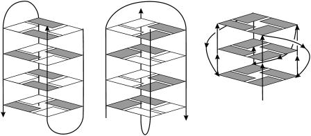
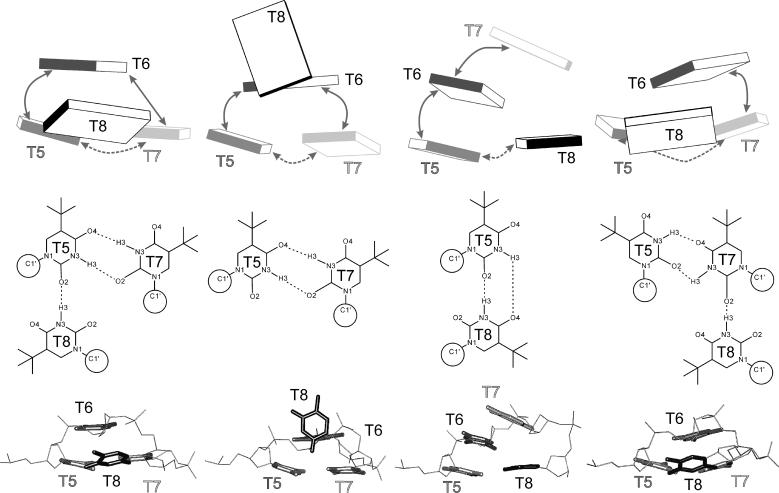
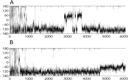
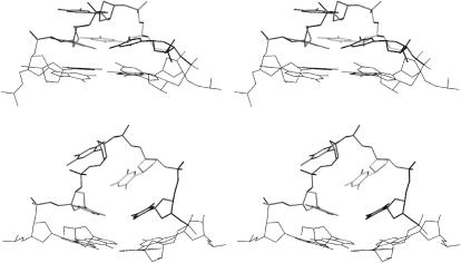
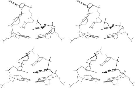
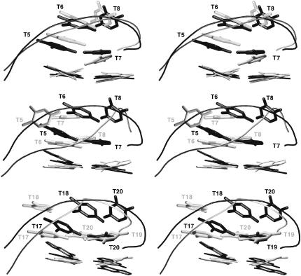
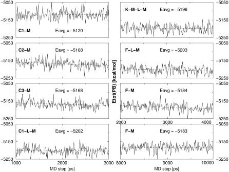
A computational analysis of d(GGGGTTTTGGGG)(2) guanine quadruplexes containing either lateral or diagonal four-thymidine loops was carried out using molecular dynamics (MD) simulations in explicit solvent, locally enhanced sampling (LES) simulations, systematic conformational search, and free energy molecular-mechanics, Poisson Boltzmann, surface area (MM-PBSA) calculations with explicit inclusion of structural monovalent cations. The study provides, within the approximations of the applied all-atom additive force field, a qualitatively complete analysis of the available loop conformational space. The results are independent of the starting structures. Major conformational transitions not seen in conventional MD simulations are observed when LES is applied. The favored LES structures consistently provide lower free energies (as estimated by molecular-mechanics, Poisson Boltzmann, surface area) than other structures. Unfortunately, the predicted optimal structure for the diagonal loop arrangement differs substantially from the atomic resolution experiments. This result is attributed to force field deficiencies, such as the potential misbalance between solute-cation and solvent-cation terms. The MD simulations are unable to maintain the stable coordination of the monovalent cations inside the diagonal loops as reported in a recent x-ray study. The optimal diagonal and lateral loop arrangements appear to be close in energy although a proper inclusion of the loop monovalent cations could stabilize the diagonal architecture.
Figures
Similar articles
-
Structural dynamics and cation interactions of DNA quadruplex molecules containing mixed guanine/cytosine quartets revealed by large-scale MD simulations.J Am Chem Soc. 2001 Apr 11;123(14):3295-307. doi: 10.1021/ja002656y. J Am Chem Soc. 2001. PMID: 11457065
-
Single Stranded Loops of Quadruplex DNA As Key Benchmark for Testing Nucleic Acids Force Fields.J Chem Theory Comput. 2009 Sep 8;5(9):2514-30. doi: 10.1021/ct900200k. Epub 2009 Aug 19. J Chem Theory Comput. 2009. PMID: 26616629
-
A double chain reversal loop and two diagonal loops define the architecture of a unimolecular DNA quadruplex containing a pair of stacked G(syn)-G(syn)-G(anti)-G(anti) tetrads flanked by a G-(T-T) Triad and a T-T-T triple.J Mol Biol. 2001 Jun 29;310(1):181-94. doi: 10.1006/jmbi.2001.4759. J Mol Biol. 2001. PMID: 11419945
-
Molecular dynamic simulations of environment and sequence dependent DNA conformations: the development of the BMS nucleic acid force field and comparison with experimental results.J Biomol Struct Dyn. 1998 Dec;16(3):487-509. doi: 10.1080/07391102.1998.10508265. J Biomol Struct Dyn. 1998. PMID: 10052609 Review.
-
Frontiers in molecular dynamics simulations of DNA.Acc Chem Res. 2012 Feb 21;45(2):196-205. doi: 10.1021/ar2001217. Epub 2011 Aug 10. Acc Chem Res. 2012. PMID: 21830782 Review.
Cited by
-
Identification of mixed di-cation forms of G-quadruplex in solution.Nucleic Acids Res. 2005 Jun 28;33(11):3691-7. doi: 10.1093/nar/gki690. Print 2005. Nucleic Acids Res. 2005. PMID: 15985684 Free PMC article.
-
Origin of Ion Specificity of Telomeric DNA G-Quadruplexes Investigated by Free-Energy Simulations.Biophys J. 2017 Jun 6;112(11):2280-2290. doi: 10.1016/j.bpj.2017.04.036. Biophys J. 2017. PMID: 28591601 Free PMC article.
-
Loss of G-A base pairs is insufficient for achieving a large opening of U4 snRNA K-turn motif.Nucleic Acids Res. 2005 Jun 13;33(10):3435-46. doi: 10.1093/nar/gki664. Print 2005. Nucleic Acids Res. 2005. PMID: 15956103 Free PMC article.
-
Toward Improved Description of DNA Backbone: Revisiting Epsilon and Zeta Torsion Force Field Parameters.J Chem Theory Comput. 2013 May 14;9(5):2339-2354. doi: 10.1021/ct400154j. J Chem Theory Comput. 2013. PMID: 24058302 Free PMC article.
-
Ion Binding Properties and Dynamics of the bcl-2 G-Quadruplex Using a Polarizable Force Field.J Chem Inf Model. 2020 Dec 28;60(12):6476-6488. doi: 10.1021/acs.jcim.0c01064. Epub 2020 Dec 2. J Chem Inf Model. 2020. PMID: 33264004 Free PMC article.
References
-
- Balagurumoorthy, P., and S. K. Brahmachari. 1994. Structure and stability of human telomeric sequence. J. Biol. Chem. 269:21858–21869. - PubMed
-
- Berendsen, H. J. C., J. P. M. Postma, W. F. Vangunsteren, A. Dinola, and J. R. Haak. 1984. Molecular-dynamics with coupling to an external bath. J. Chem. Phys. 81:3684–3690.
-
- Beveridge, D. L., and K. J. McConnell. 2000. Nucleic acids: theory and computer simulation, Y2K. Curr. Opin. Struct. Biol. 10:182–196. - PubMed
-
- Bouaziz, S., A. Kettani, and D. J. Patel. 1998. A K cation-induced conformational switch within a loop spanning segment of a DNA quadruplex containing G-G-G-C repeats. J. Mol. Biol. 282:637–652. - PubMed
Publication types
MeSH terms
Substances
LinkOut - more resources
Full Text Sources
Other Literature Sources
