. 2004 Jul;87(1):419-29.
doi: 10.1529/biophysj.104.040360.

A new method for the reconstitution of membrane proteins into giant unilamellar vesicles

Affiliations

A new method for the reconstitution of membrane proteins into giant unilamellar vesicles

Philippe Girard et al. Biophys J. 2004 Jul.

Erratum in

  • Biophys J. 2004 Sep;87(3):2098

Abstract

In this work, we have investigated a new and general method for the reconstitution of membrane proteins into giant unilamellar vesicles (GUVs). We have analyzed systematically the reconstitution of two radically different membrane proteins, the sarcoplasmic reticulum Ca(2+)-ATPase and the H(+) pump bacteriorhodopsin. In a first step, our method involved a detergent-mediated reconstitution of solubilized membrane proteins into proteoliposomes of 0.1-0.2 microm in size. In a second step, these preformed proteoliposomes were partially dried under controlled humidity followed, in a third step, by electroswelling of the partially dried film to give GUVs. The physical characteristics of GUVs were analyzed in terms of morphology, size, and lamellarity using phase-contrast and differential interference contrast microscopy. The reconstitution process was further characterized by analyzing protein incorporation and biological activity. Both membrane proteins could be homogeneously incorporated into GUVs at lipid/protein ratios ranging from 5 to 40 (w/w). After reconstitution, both proteins retained their biological activity as demonstrated by H(+) or Ca(2+) pumping driven by bacteriorhodopsin or Ca(2+)-ATPase, respectively. This constitutes an efficient new method of reconstitution, leading to the production of large unilamellar membrane protein-containing vesicles of more than 20 microm in diameter, which should prove useful for functional and structural studies through the use of optical microscopy, optical tweezers, microelectrodes, or atomic force microscopy.


Figures
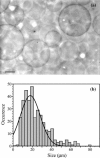
FIGURE 1
FIGURE 1
Size distribution of reconstituted GUVs. (a) Phase-contrast image of EYPC/EYPA (9:1) GUVs containing Ca2+-ATPase at a lipid/protein ratio of 6.5 w/w. Scale bar = 10 μm. (b) Size distribution of the Ca2+-ATPase reconstituted GUVs. Vesicles with a diameter lower than 5 μm were not counted.
FIGURE 2
FIGURE 2
Unilamellarity of reconstituted GUVs using a fluorescent quenching assay. Giant vesicles were prepared with a lipid mixture containing EYPC/NBD-C12-HPC (99.5:0.5 mol/mol) at a lipid/Ca2+-ATPase ratio of 6.5 w/w. (a) Unilamellarity of the GUVs was checked by measuring the distribution of fluorescent lipids between inner and outer monolayers as described in Materials and Methods. Addition of sodium hydrosulfite to reconstituted GUVs (arrow 1) induced a rapid quenching of the fluorescent lipids present in the outer lipid layer. Solubilization of the vesicles by an excess of Triton X-100 (arrow 2) induced a second decrease in fluorescence due to the quenching of the inner lipids. The I1/I2. ratio is an index of the unilamellarity of the vesicles and should be equal to 0.5 for a perfect unilamellarity. The value of 0.6 found for reconstituted GUVs could be accounted for by the presence of small vesicles (b) or other lipidic structures (arrows in c) that were sometimes visible encapsulated by the GUVs. Scale bars of phase-contrast images correspond to 5 μm.
FIGURE 3
FIGURE 3
Unilamellarity of reconstituted GUVs using elastic bending measurement. Giant vesicles were prepared in EYPC/EYPA (9:1) at a lipid/Ca2+-ATPase ratio of 6.5 w/w. (a) Microscopy image of a reconstituted GUV aspirated with a suction pressure by a micropipette and observed with a 63× differential interference contrast (DIC) oil immersion objective. DP, pipette diameter; Dv, vesicle diameter; and Lp, projection length inside the micropipette. Scale bar = 5 μm. (b) Plot of membrane tension versus the area dilation of a reconstituted vesicle. The solid curve is fitted with an exponential function based on a bending rigidity modulus κ = (9.3 ± 1.1) × kBT. Histograms of elastic bending moduli from micropipette aspiration experiments on 80 Ca2+-ATPase reconstituted GUVs (c) and on 100 pure lipidic GUVs (d).
FIGURE 4
FIGURE 4
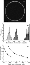
Homogeneity of Ca2+-ATPase incorporation in GUVs. (a) Fluorescence intensity image (false color representation) of a GUV reconstituted at a SOPC/FITC labeled Ca2+-ATPase ratio of 6.5 w/w. Scale bar = 5 μm. (b) Fluorescence distribution of 50 GUVs reconstituted at lipid/Ca2+-ATPase ratios of 3.5 (dark shading), 6.5 (light shading), and 12.5 w/w (open). A normal distribution function has been superimposed for each ratio giving the mean and standard deviation. (c) The average of the normalized fluorescence intensity, obtained in b, is reported as a function of the lipid to Ca2+-ATPase (▪) or lipid/BR (○) ratios.
FIGURE 5
FIGURE 5
Homogeneity of BR incorporation in GUVs. (a) Confocal image of FITC-labeled BR reconstituted into SOPC vesicles. The fluorescence is homogenously distributed in the membrane of the vesicle. (b) Epifluorescence videomicroscopy frame (courtesy of Dr. J.-B. Manneville, Unité Mixte de Recherche 144 Centre National de la Recherche Scientifique/Institut Curie, Paris) of FITC-labeled BR reconstituted into SOPC vesicles according to the protocol of Manneville et al. (2001). The fluorescence of the membrane is not homogenous suggesting the presence of BR aggregates. Scale bars = 5 μm.
FIGURE 6
FIGURE 6
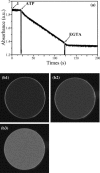
Activity of Ca2+-ATPase into GUVs. Giant vesicles were prepared in EYPC/EYPA (9:1) at a lipid/Ca2+-ATPase ratio of 6.5 w/w. (a) ATPase activity was performed at 25°C as described in Materials and Methods. The reconstituted GUVs were transferred to a cuvette and solubilized with C12E8 (arrow 1). The reaction was started by the addition of ATP and stopped with EGTA. (b) Calcium pumping activity. Confocal images of a Ca2+-ATPase GUV (30 μm in diameter) reconstituted in EYPC/EYPA/NBD-C12-HPC s and encapsulating Fluo-5N. After ATP addition, the fluorescence intensity in the interior of the vesicles increases as a function of time. (b1) t = 5 min; (b2) t = 15 min; and (b3) t = 25 min.
FIGURE 7
FIGURE 7
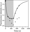
Proton pumping activity of BR into GUVs. Light-induced fluorescence responses of pyranine trapped inside giant vesicles. Giant vesicles containing pyranine were prepared in SOPC at a lipid/BR ratio of 7 (w/w) and analyzed by confocal microscopy. In the shaded part, BR is illuminated and pumps H+. In the open part, the illumination is stopped, and the pH reequilibrates. ▵ correspond to the direct measured variation of fluorescence intensities. ○ correspond to the fluorescence intensities corrected for the photobleaching. • correspond to the extrapolated initial fluorescence intensities since the vesicles were already illuminated in the confocal microscope during the installation of the sample and selection of the vesicle, leading to a pumping of protons before the fluorescence measurement.

References

    1. Abdelghani-Jacquin, C., A. Abdelghani, M. Kantlehner, and E. Sackmann. 2002. Decorated surfaces by biofunctionalized gold beads: application to cell adhesion studies. Eur. Biophys. J. 2:102–110. - PubMed
    1. Ajouz, B., C. Berrier, M. Besnard, B. Martinac, and A. Ghazi. 2000. Contributions of the different extramembranous domains of the mechanosensitive ion channel MscL to its response to membrane tension. J. Biol. Chem. 275:1015–1022. - PubMed
    1. Alberts, B., D. Bray, J. Lewis, M. Raff, K. Roberts, and J. D. Watson. 1994. Molecular Biology of the Cell, 3rd ed. Garland Publishing, New York.
    1. Angelova, M. I. 2000. Liposome electroformation. In Giant Vesicles. P. L. Luisi and P. Walde, editors. John Wiley and Sons, Chichester, UK. 27–36.
    1. Angelova, M. I., N. Hristova, and I. Tsoneva. 1999. DNA-induced endocytosis upon local microinjection to giant unilamellar cationic vesicles. Eur. Biophys. J. 28:142–150. - PubMed

Publication types

MeSH terms