. 2004 Jul;87(1):562-73.
doi: 10.1529/biophysj.103.037333.

The unfolding action of GroEL on a protein substrate

Affiliations

The unfolding action of GroEL on a protein substrate

Arjan van der Vaart et al. Biophys J. 2004 Jul.

Abstract

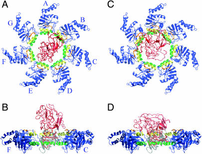
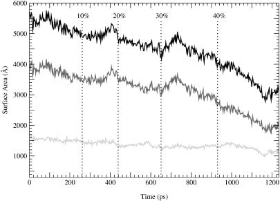
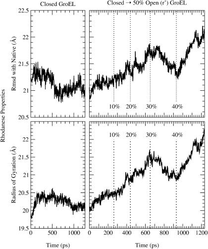
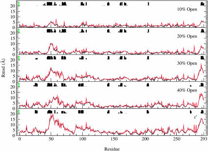
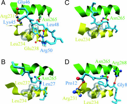
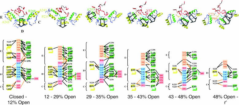
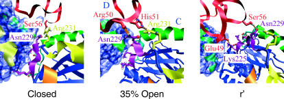
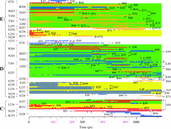
A molecular dynamics simulation of the active unfolding of denatured rhodanese by the chaperone GroEL is presented. The compact denatured protein is bound initially to the cis cavity and forms stable contacts with several of the subunits. As the cis ring apical domains of GroEL undergo the transition from the closed to the more open (ATP-bound) state, they exert a force on rhodanese that leads to the increased unfolding of certain loops. The contacts between GroEL and rhodanese are analyzed and their variation during the GroEL transition is shown. The major contacts, which give rise to the stretching force, are found to be similar to those observed in crystal structures of peptides bound to the apical domains. The results of the simulation show that multidomain interactions play an essential role, in accord with experiments. Implications of the results for mutation experiments and for the action of GroEL are discussed.


Figures

FIGURE 1
FIGURE 1
Snapshots of the initial configurations of rhodanese. A and B show the starting configuration for the study of denatured rhodanese and closed-state GroEL; there are no contacts between GroEL and rhodanese in this configuration. C and D show the starting configurations for the TMD simulation; these are the endpoints of the closed-state simulation. The overall motion of rhodanese during the closed-state simulation is a translation by 10.7 Å and a 45.9° rotation. The H helices of GroEL are colored yellow, the I helices are green, the loop formed by residues 310–315 is orange, and the rest of the GroEL apical domains are blue. Rhodanese is shown in red. A and C show the top views, in which the A subunit is at the 12-o'clock position and the other subunits (B–G) follow in a clockwise fashion as indicated. B and D show the side views, obtained by a 90° rotation. For clarity, subunits D and E have been removed in B and D, and subunit A is colored gray. Figs. 1, 2, and 6–8 were prepared with VMD (Humphrey et al., 1996).
FIGURE 2
FIGURE 2
Snapshots during the unfolding simulation; for the description of the different states, see Methods and text. The H helices of GroEL are colored yellow, the I helices are green, the loops formed by residues 310–315 are orange, and the rest of GroEL is blue. GroEL is viewed from the top, looking down into the cis cavity, as in Fig. 1. Subunit A is at the 12-o'clock position, the other subunits (B–G) follow in a clockwise fashion (see Fig. 1). Rhodanese is shown in red, except for the loop consisting of residues 45–50, which is in light blue.
FIGURE 3
FIGURE 3
Contact area between GroEL and rhodanese during the GroEL closed-to-r′ transition. The black line corresponds to the total contact area, the dark gray line to the hydrophobic portion of the contact area, and the light gray line to the polar portion of the contact area.
FIGURE 4
FIGURE 4
Rhodanese properties during the simulation. The top panels show the RMSD from the native state, and the bottom panels show the radius of gyration. The left panels correspond to rhodanese bound to the closed state of GroEL, whereas the right panels correspond to the closed-to-r′ state transition.
FIGURE 5
FIGURE 5
The RMSD per residue for rhodanese during the closed-to-r′ state transition of GroEL. The RMSD is with respect to the closed-state bound structure. Black lines show the backbone RMSD, red lines the RMSD for the entire residue. Contacts between rhodanese and GroEL are indicated by the histograms. The height of the histograms represents the lifetime of these contacts during each stage of the transition; these lifetimes were measured between the closed and 10% open state for the top plot, between the 10% and 20% open state for the 20% open plot, etc. The maximum possible lifetimes are indicated by the green bars on the left; it is evident that many contacts are present during the entire transition (see text).
FIGURE 6
FIGURE 6
Binding to closed-state GroEL in simulation and experiments. The peptides are shown in light blue, the GroEL H helix is yellow, and the GroEL I helix is shown in green. The hydrophobic pocket formed by Leu-234 and Leu-237 of the H helix is shown by the gray surface. Hydrogen bonds between the peptide and GroEL are indicated by the orange dotted lines; intramolecular hydrogen bonds in the peptides are indicated by blue dotted lines. (A) The starting TMD configuration for rhodanese residues 45–50. Hydrogen bonds are formed between rhodanese Lys-45 and GroEL Arg-231, rhodanese Glu-46 and GroEL Arg-231, rhodanese Tyr-47 and Asn-265, and rhodanese Arg-50 and GroEL Glu-238. Intramolecular hydrogen bonds exist between rhodanese Tyr-47 and Arg-50 and between rhodanese Leu-48 and Arg-50. For clarity, the hydrogen bond between Glu-46 and Arg-268 of the E subunit is excluded from the figure. The hydrophobic pocket is occupied by Tyr-47. (B) GroES residues 24–30 binding to GroEL (Xu et al., 1997). There is a hydrogen bond between GroES Leu-27 and GroEL Asn-265; the hydrophobic pocket is occupied by Val-26. (C) Residues 184-189 of the N-terminal extension binding to GroEL (Buckle et al., 1997). There is a hydrogen bond between Val-186 of the N-terminal extension and Asn-265 of GroEL. The hydrophobic pocket is occupied by Leu-185. (D) Residues 6–12 of the SBP peptide binding to GroEL, taken from chain F and B of the protein data bank structure 1DKD (Chen and Sigler, 1999). Hydrogen bonds are formed between SBP Gly8 and GroEL Arg-268, SBP Leu-10, and GroEL Asn-265, and SBP Pro-12 and GroEL Arg-231. The hydrophobic pocket is occupied by Phe-9.
FIGURE 7
FIGURE 7
Stretching and interactions for the 42–70 loop of rhodanese. The H helices of GroEL (residues 234–243) are colored yellow, the I helices (residues 257–268) are green, and the rest of GroEL is blue. The symbols C, D, and E refer to the different GroEL subunits. GroEL is viewed from the top of subunit D, looking down into the cis cavity. The viewing angle is the same for all snapshots. Rhodanese residues 45–50 are light blue, residues 67–72 are orange; the other rhodanese residues are shown in red. The direction of the stretching force is indicated by the black arrows. The diagrams below show the interactions between these rhodanese residues and GroEL during part of the transition. Hydrogen bonding is indicated by the orange lines, heavy atom contacts within 4.0 Å, which are mainly van der Waals contacts, are shown by the black lines. The diagrams show all interactions that are present for 10 ps or more during the entire interval; not all contacts and hydrogen bonds are present at every instant of the interval. In the diagrams the H helix is shown on a yellow background, the I helix on a green background, and other GroEL residues are shown on a white background. Residue Arg-231 of GroEL is shown on a yellow background to indicate the closeness of this residue to the H helix. The coloring of rhodanese is identical to the structures above. Binding of rhodanese parallel to the H helix is indicated by a yellow bar (left), binding of rhodanese parallel to the I helix by the green bar (right and left). The presence of both bars for certain residues (e.g., Glu-46, Tyr-47) indicate that this residue was bound in the cleft formed by the H and I helices; this happens in the closed to 12% open and the 12% to 29% open intervals.
FIGURE 8
FIGURE 8
The interface between the apical domains of the C and D subunit (see text). The H helices of GroEL are colored yellow, the I helices are green, loop 310–315 is orange, loop 225–230 is light blue, and loop 252–256 is purple; the rest of GroEL is blue. Subunit C is shown as a ribbon diagram with the polar and charged side chains of loops 225–230 and 252–256 of subunit C as stick models; Arg-231 is shown as a yellow stick model to indicate the closeness of this residue to the H helix (analogous to Fig. 7). For subunit D the solvent-accessible surface is shown in blue. Rhodanese is shown in red, hydrogen bonds between GroEL residues 225–231 and rhodanese are indicated by the orange dotted lines. GroEL is viewed from the top; the viewing angle is identical to that of Fig. 2.
FIGURE 9
FIGURE 9
Heavy atom contacts within 4.0 Å between rhodanese residues 42–72 and GroEL; the conventions (labels and colors) are the same as in the other figures. This figure should be used in conjunction with Fig. 7. GroEL residues of subunits C, D, and E are shown on the left; the vertical bars group contacts with a given residue. The H helices and Arg-231 are indicated by the yellow background, the I helices by the green background; other GroEL residues are shown on a white background. Rhodanese residues are indicated by the arrows; residues 45–50 are shown in blue, residues 67–72 in orange, and the other residues in red.

Similar articles

Cited by

References

    1. Anfinsen, C. 1973. Principles that govern the folding of protein chains. Science. 181:223–230. - PubMed
    1. Betancourt, M., and D. Thirumalai. 1999. Exploring the kinetic requirements for enhancement of protein folding rates in the GroEL cavity. J. Mol. Biol. 287:627–644. - PubMed
    1. Böckmann, R., and H. Grubmüller. 2002. Nanoseconds molecular dynamics simulation of primary mechanical energy transfer steps in F1-ATP synthase. Nat. Struct. Biol. 9:198–202. - PubMed
    1. Boisvert, D., J. Wang, Z. Otwinowski, A. Horwich, and P. Sigler. 1996. The 2.4 Å crystal structure of the bacterial chaperonin GroEL complexed with ATPγS. Nat. Struct. Biol. 3:170–177. - PubMed
    1. Brinker, A., G. Pfeifer, M. Kerner, D. Naylor, F. Hartl, and M. Hayer-Hartl. 2001. Dual function of protein confinement in chaperonin-assisted protein folding. Cell. 107:223–233. - PubMed

Publication types

MeSH terms