

doi: 10.1083/jcb.200406019.
What's in a picture? The temptation of image manipulation
- PMID: 15240566
- PMCID: PMC2172141
- DOI: 10.1083/jcb.200406019
Item in Clipboard
What's in a picture? The temptation of image manipulation
J Cell Biol.
.
No abstract available
Figures
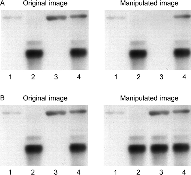
Gross manipulation of blots. (A) Example of a band deleted from the original data (lane 3). (B) Example of a band added to the original data (lane 3).
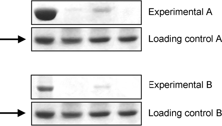
Gross manipulation of blots. Example of a duplicated panel (arrows).
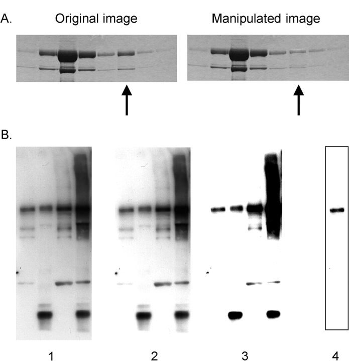
Manipulation of blots: brightness and contrast adjustments. (A) Adjusting the intensity of a single band (arrow). B) Adjustments of contrast. Images 1, 2, and 3 show sequentially more severe adjustments of contrast. Although the adjustment from 1 to 2 is acceptable because it does not obscure any of the bands, the adjustment from 2 to 3 is unacceptable because several bands are eliminated. Cutting out a strip of a blot with the contrast adjusted provides the false impression of a very clean result (image 4 was derived from a heavily adjusted version of the left lane of image 1). For a more detailed discussion of “gel slicing and dicing,” see Nature Cell Biology editorial (2).
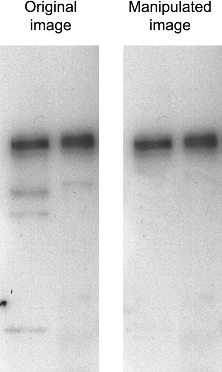
Manipulation of blots: cleaning up background. The Photoshop “Rubber Stamp” tool has been used in the manipulated image to clean up the background in the original data. Close inspection of the image reveals a repeating pattern in the left lane of the manipulated image, indicating that such a tool has been used.
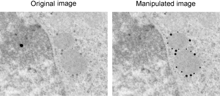
Misrepresentation of immunogold data. The gold particles, which were actually present in the original (left), have been enhanced in the manipulated image (right). Note also that the background dot in the original data has been removed in the manipulated image.
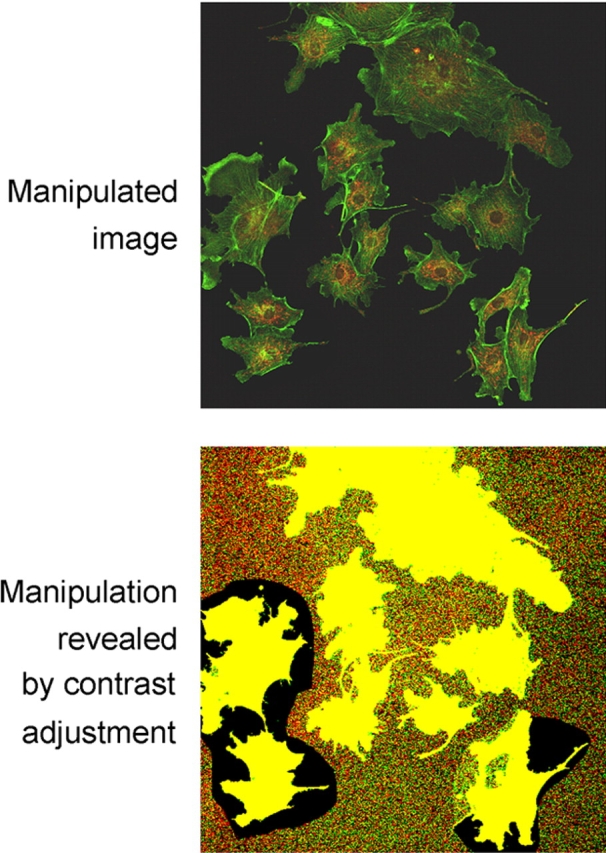
Misrepresentation of image data. Cells from various fields have been juxtaposed in a single image, giving the impression that they were present in the same microscope field. A manipulated panel is shown at the top. The same panel, with the contrast adjusted by us to reveal the manipulation, is shown at the bottom.
References
Publication types
MeSH terms
LinkOut - more resources
Full Text Sources
Other Literature Sources
