. 2004 Jul 5;166(1):37-47.
doi: 10.1083/jcb.200311021.

SOX9 is an intestine crypt transcription factor, is regulated by the Wnt pathway, and represses the CDX2 and MUC2 genes

Affiliations

SOX9 is an intestine crypt transcription factor, is regulated by the Wnt pathway, and represses the CDX2 and MUC2 genes

Philippe Blache et al. J Cell Biol. .

Abstract

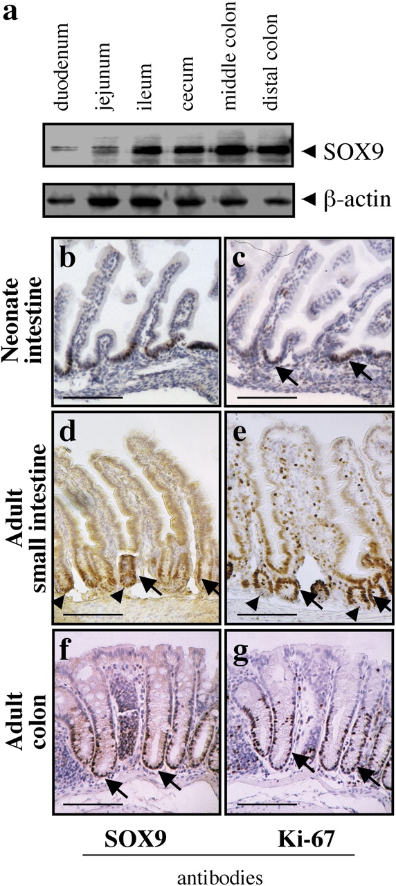
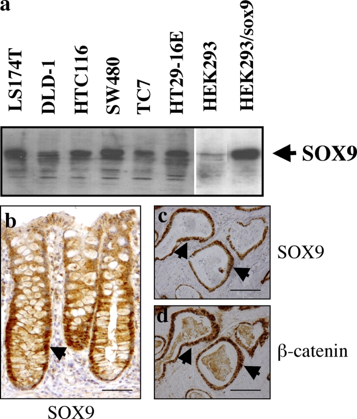
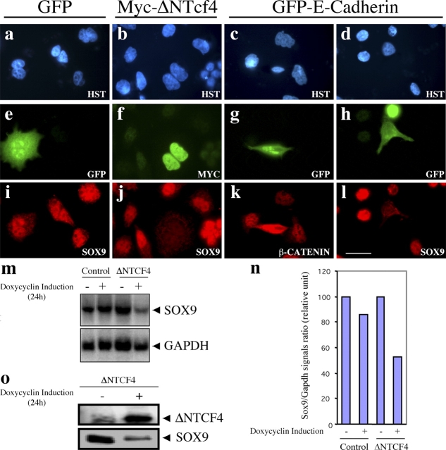
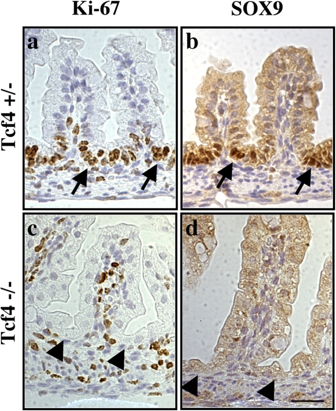
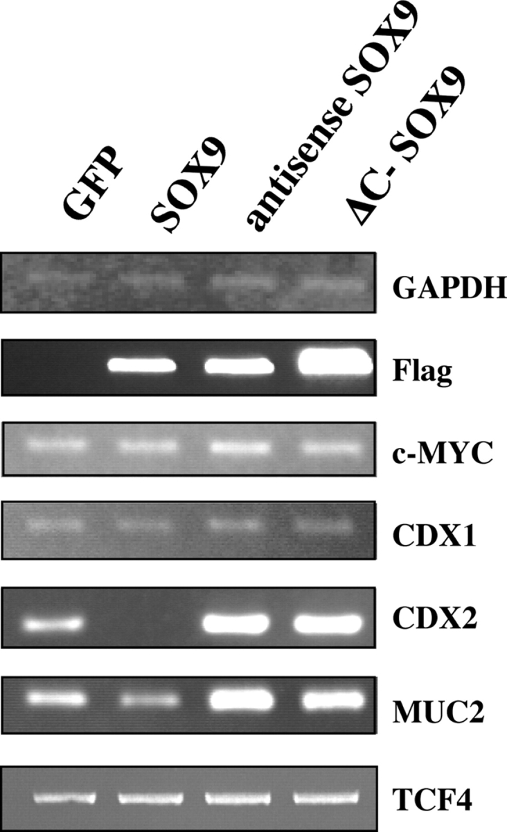
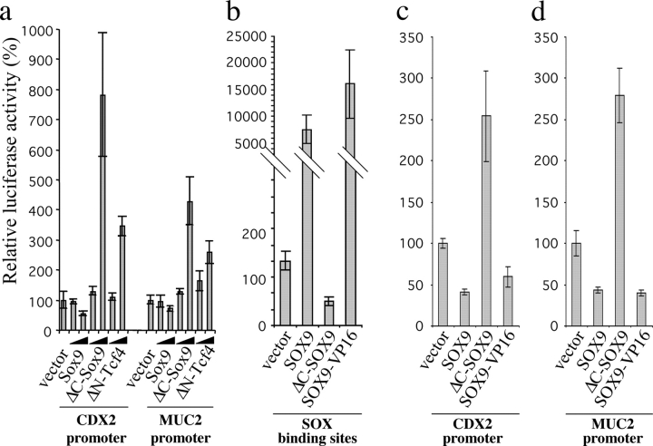
TCF and SOX proteins belong to the high mobility group box transcription factor family. Whereas TCFs, the transcriptional effectors of the Wnt pathway, have been widely implicated in the development, homeostasis and disease of the intestine epithelium, little is known about the function of the SOX proteins in this tissue. Here, we identified SOX9 in a SOX expression screening in the mouse fetal intestine. We report that the SOX9 protein is expressed in the intestinal epithelium in a pattern characteristic of Wnt targets. We provide in vitro and in vivo evidence that a bipartite beta-catenin/TCF4 transcription factor, the effector of the Wnt signaling pathway, is required for SOX9 expression in epithelial cells. Finally, in colon epithelium-derived cells, SOX9 transcriptionally represses the CDX2 and MUC2 genes, normally expressed in the mature villus cells of the intestinal epithelium, and may therefore contribute to the Wnt-dependent maintenance of a progenitor cell phenotype.


Figures

Figure 1.
Figure 1.
SOX9 is expressed in the proliferative compartment of the epithelium. (a) Western blot with mouse tissue samples representative of the overall length of the intestine (5 μg protein/lane). Antibodies used are indicated. (b) Immunohistochemistry with neonate and adult intestine samples. In the neonate mouse, the architecture of the intestine is not complete, crypts are not formed and the proliferative, Ki-67 positive, compartment is restricted to the intervillus region (top). In the adult small intestine, crypts are invaginated between the villi in the underlying mesenchyme (middle). In the adult colon, crypts contain more cells, villi are absent and the differentiated compartment constitutes the flat luminal surface of the epithelium. Cells constituting the proliferative compartment are positive for both SOX9 and Ki-67 (arrows). Arrowheads point at Paneth cells, positive for SOX9 and negative for Ki-67. Bars: (b–e) 50 μm; (f and g) 40 μm.
Figure 2.
Figure 2.
SOX9 is expressed in various colon carcinoma cell lines. (a) Western blot with cell extracts from a panel of human colon carcinoma cell lines (1 μg/lane) and the nonintestinal epithelial cell line HEK293. Endogenous SOX9 protein was detected in all colon carcinoma–derived cell lines but not in HEK293. HEK293 cells, transiently transfected with SOX9, were used as a positive control. (b–d). Immunohistochemistry with sections of human colon. (b) SOX9 staining in healthy colon epithelium; (c) SOX9 staining in adenocarcinomatous colon; (d) β-catenin staining in adenocarcinomatous colon. Arrows point at cells expressing the indicated proteins. Bars: (b) 50 μm; (c and d) 80 μm.
Figure 3.
Figure 3.
The Wnt–β-catenin–Tcf pathway is required for SOX9 expression in colon epithelial cells. (a–l) Immunofluorescence in transiently transfected LS174T colon carcinoma cells, fixed 36 h after transfection. (a–d) Hoechst nuclear staining. (e) GFP. (f) Detection of exogenous Myc-tagged ΔNTCF4 in transfected cells with an anti-Myc antibody. (g and h) GFP shows the perinuclear accumulation of the GFP–cyt-E-cadherin fusion in transfected cells. (i) Identical SOX9 staining in GFP-transfected and nontransfected cells. (j and l) SOX9 staining is absent in cells transfected with the Wnt pathway interfering constructs ΔNTCF4 and GFP–cyt-E-cadherin. (k) β-Catenin is efficiently sequestered by GFP–cyt-E-cadherin, as shown by their identical distribution in transfected cells. The type of staining is indicated on each panel. (m) Northern blot. SOX9 mRNA detection in LS174T colon carcinoma cells stably transfected with an inducible ΔNTCF4 construct. Time of doxycyclin induction is indicated for both the control and the ΔNTCF4 expressing cell line. A GAPDH probe was used as a loading control. (n) Phosphorimager quantification of the Northern blot signals. (o) Western blot. Detection of the SOX9 protein in the ΔNTCF4 stably transfected LS174T cells with or without doxycycline induction. Bar, 15 μm.
Figure 4.
Figure 4.
Absence of the SOX9 protein in the proliferative compartment of TCF4-deficient neonate mouse. (a and b) TCF4 heterozygous mice are healthy and fertile. The actively proliferating intervillus cells are Ki-67 positive and express abundant nuclear SOX9 protein (arrows). (c and d) In TCF4 null mice, no Ki-67 positive proliferating cells can be found in the intervillus region (arrowheads), and SOX9 expression is completely abrogated. Bar, 5 μm.
Figure 5.
Figure 5.
SOX9 represses intestine epithelium differentiation genes. LS174T cells were transiently transfected with GFP, Flag-SOX9 fusion, Flag-SOX9 antisense, or Flag-COOH–truncated SOX9 (ΔC-Sox9) constructs, and the expression of the indicated putative target genes was monitored by RT-PCR. The primers used are listed in Table S1.
Figure 6.
Figure 6.
SOX9 negatively regulates the promoters of the CDX2 and MUC2 genes. LS174T cells were cotransfected, in triplicate, with the indicated expression constructs (10 and 100 ng) together with CDX2-luciferase, MUC2-luciferase and SOX-luciferase reporters (500 ng) and relative luciferase activities were measured 36 h after transfection. The reporter activity in mock-transfected cells was arbitrarily set to 100. (a) CDX2 and MUC2 promoters regulation by SOX9; (b–d) SOX9-VP16 recapitulates the transcription regulation properties of the wild-type SOX9 on SOX-luciferase (b), CDX2-luciferase (c), and MUC2-luciferase constructs (d). The histograms represent mean values of triplicate experiments and SDs are shown with error bars.
Figure 7.
Figure 7.
SOX9 represses CDX2 and MUC2 in xenograft-derived tumors. Sections from tumors resulting from xenografts of HT29-16E-SOX9 cells, stained with antibodies against the Flag tag, the CDX2 and MUC2 proteins. Antibodies used for staining are indicated. The expression of the Flag-SOX9 construct is detected uniquely in the nucleus of tumors from doxycycline-fed mice. Bar, 130 μm.

References

    1. Akiyama, H., M.C. Chaboissier, J.F. Martin, A. Schedl, and B. de Crombrugghe. 2002. The transcription factor Sox9 has essential roles in successive steps of the chondrocyte differentiation pathway and is required for expression of Sox5 and Sox6. Genes Dev. 16:2813–2828. - PMC - PubMed
    1. Aoki, K., Y. Tamai, S. Horiike, M. Oshima, and M.M. Taketo. 2003. Colonic polyposis caused by mTOR-mediated chromosomal instability in Apc+/Δ716 Cdx2+/− compound mutant mice. Nat. Genet. 35:323–330. - PubMed
    1. Avilion, A.A., S.K. Nicolis, L.H. Pevny, L. Perez, N. Vivian, and R. Lovell-Badge. 2003. Multipotent cell lineages in early mouse development depend on SOX2 function. Genes Dev. 17:126–140. - PMC - PubMed
    1. Batlle, E., J.T. Henderson, H. Beghtel, M.M.W. van den Born, E. Sancho, G. Huls, J. Meeldijk, J. Robertson, M. van de Wetering, T. Pawson, and H. Clevers. 2002. β-Catenin and TCF mediate cell positioning in the intestinal epithelium by controlling the expression of EphB/EphrinB. Cell. 111:251–263. - PubMed
    1. Bell, D.M., K.K.H. Leung, S.C. Wheatley, L.J. Ng, S. Zhou, K.W. Ling, M.H. Sham, P. Koopman, P.P.L. Tam, and K.S.E. Cheah. 1997. SOX9 directly regulates the type-II collagen gene. Nat. Genet. 16:174–178. - PubMed

Publication types

MeSH terms