. 2004 Jul 5;166(1):97-109.
doi: 10.1083/jcb.200401150.

Endorepellin causes endothelial cell disassembly of actin cytoskeleton and focal adhesions through alpha2beta1 integrin

Affiliations

Endorepellin causes endothelial cell disassembly of actin cytoskeleton and focal adhesions through alpha2beta1 integrin

Gregory Bix et al. J Cell Biol. .

Erratum in

  • J Cell Biol. 2013 May 13;201(4):641

Abstract

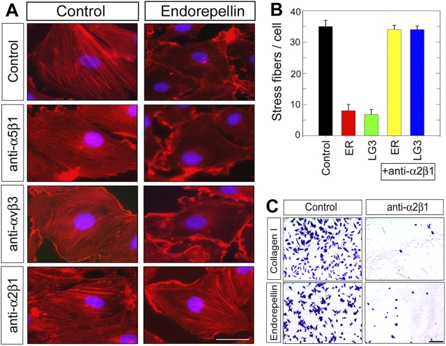
Endorepellin, the COOH-terminal domain of the heparan sulfate proteoglycan perlecan, inhibits several aspects of angiogenesis. We provide evidence for a novel biological axis that links a soluble fragment of perlecan protein core to the major cell surface receptor for collagen I, alpha2beta1 integrin, and provide an initial investigation of the intracellular signaling events that lead to endorepellin antiangiogenic activity. The interaction between endorepellin and alpha2beta1 integrin triggers a unique signaling pathway that causes an increase in the second messenger cAMP; activation of two proximal kinases, protein kinase A and focal adhesion kinase; transient activation of p38 mitogen-activated protein kinase and heat shock protein 27, followed by a rapid down-regulation of the latter two proteins; and ultimately disassembly of actin stress fibers and focal adhesions. The end result is a profound block of endothelial cell migration and angiogenesis. Because perlecan is present in both endothelial and smooth muscle cell basement membranes, proteolytic activity during the initial stages of angiogenesis could liberate antiangiogenic fragments from blood vessels' walls, including endorepellin.


Figures

Figure 1.
Figure 1.
ER and its COOH-terminal LG3 module disrupt capillary morphogenesis in Matrigel. (A) Purification of ER and two fragments encompassing the LG1/LG2 and LG3 domains, respectively, as schematically shown in the right margin. The picture represents a Coomassie-stained SDS-PAGE of purified recombinant proteins. (B–D) Light micrographs of normal breast (B), breast carcinoma (C), and astrocytoma (D) showing specific labeling of blood vessels using the anti-ER antibody. (E–G) Capillary morphogenesis after coculture of 293-EBNA cells (within Matrigel) and HUVECs (on top of Matrigel) as indicated. (H–J) Immunoblotting of conditioned media from the corresponding cocultures shown in E–G. Notice the presence of LG3 and ER (arrow) in the coculture with the LG3- and ER-secreting 293-EBNA cells. The nonspecific bands derived from serum proteins are labeled by asterisks. (K–N) Inhibition of HUVEC capillary morphogenesis on Matrigel by recombinant ER and LG3, but not by LG1/LG2 (150 nM, each, for 24 h). Bars, 100 μm.
Figure 2.
Figure 2.
Transduction of endothelial cells with Ad-ER prevents capillary morphogenesis in collagen overlay assays. (A) Western immunoblotting of media from various cells transduced with control adenoviral vector (Ad-Empty) or Ad-ER at the designated plaque forming unit (pfu) using anti-ER antibody (αER). About 4 × 10⁴ HUVECs and 6 × 10⁴ MECs cells were transduced with indicated amounts of Ad-Empty or Ad-ER for 48 h. 2 × 10⁴ rat breast carcinoma MTLn3 cells and 2 × 10⁴ human colon carcinoma WiDr cells were transduced with Ad-Empty or increasing amounts of Ad-ER as indicated. Conditioned media were harvested after 48 h. (B) Wells were precoated with 5 μg/cm2 human fibronectin or laminin, or with 10.5 μg/cm2 collagen I; 3 × 10⁴ HUVECs were plated and transduced with 4.8 × 10⁶ pfu of Ad-Empty or Ad-ER for 48 h. Control wells were left untransduced or treated with 150 nM ER or 50 nM LG3 for 20 min in serum-free conditions. Collagen gels were added to the cells for 24 h, to induce capillary morphogenesis. At the end of the incubation, collagen gels were removed and cells fixed and stained. Bar, 100 μm.
Figure 3.
Figure 3.
ER and its components cause disassembly of HUVEC actin stress fibers and focal adhesions. HUVECs were treated with the various recombinant proteins (25–150 nM) for 10 min as indicated, fixed, permeabilized with 0.1% Triton X-100, and stained to visualize actin stress fibers with rhodamine-phalloidin, focal adhesions with anti-vinculin mAb and appropriate FITC- conjugated secondary antibody, and counterstained with DAPI. In recovery experiments, after the 10-min exposure, ER was removed and the cells were incubated for an additional 3 h in serum-free media. Bar, 10 μm.
Figure 4.
Figure 4.
ER causes clustering and colocalization of α2β1 integrin with collapsed actin. HUVECs were treated with 150 nM ER for 10 min followed by labeling of actin (A, D, and G) or immunostaining with antibodies against α1β1, αvβ3, or α2β1 integrin (B, E, and H). Identical fields were then merged (C, F, and I). Notice the colocalization of collapsed actin stress fibers with α2β1 epitopes (G–I, arrowheads). Lower ER concentrations (50–100 nM) yielded similar results (not depicted). Bar, 10 μm.
Figure 5.
Figure 5.
ER supports cell attachment that is inhibited by α2β1 integrin blocking antibody. (A) ER action is blocked by function blocking antibody against α2β1 integrin, but not by antibodies against other integrins. The cells were processed as in the legend to Fig. 3, after 10-min incubation with saturating concentrations (10 μg/ml) of specific mAbs ± ER. Bar, 10 μm. (B) Mean number of visible actin stress fibers per cell was calculated for 100 randomly selected cells for each treatment as indicated. Error bars represent SEM. (C) Light micrographs of HUVECs attached to either collagen I or ER substrata in the presence or absence of anti-α2β1 integrin antibodies. Bar, 50 μm.
Figure 6.
Figure 6.
ER binding and activity require the α2β1 integrin. (A) Effects of ER/AP chimera on endothelial cell stress fibers. HUVECs were incubated for 10 min with medium containing ∼200 nM ER/AP, before and after heat denaturation (65°C, for 45 min). Bar, 10 μm. (B) Western immunoblotting of total cell lysates from NMuMg lacking or expressing the α2 integrin subunit, as indicated. (C) Dose-dependent binding of ER/AP-containing media to the α2+ cells. Cells were incubated for 1 h at 25°C with ER/AP media at the indicated dilutions (equivalent to 200, 100, 50, and 25 nM ER/AP), washed and processed for AP chemiluminescence and exposure to an X-ray film. (D) Saturation binding curves using an experimental strategy as in C, with the exception that the values were obtained by measuring the relative light units of total bound ER/AP and by normalizing the results on a standard curve generated with recombinant placental AP. Values represent the mean ± SEM (n = 3). (E) Scatchard analysis shows one single class of surface receptors with relative high affinity (K D = 23 ± 2 nM). (F and G) Analyses of ER and LG3 binding to the I domain of α2 integrin by SPR. About 3,000 response units (RU) of α2I domain were immobilized on a CM5 chip, and increasing concentrations (0.1–2.0 μM, from bottom to top line) of ER or LG3 were tested as indicated in a buffer containing 1 mM MgCl2. The profiles were corrected for response over a reference flow cell containing no immobilized protein. (H) Recombinant α2I but not α1I integrin domain or GST protein, blocks ER effects on HUVECs. Recombinant ER was incubated with α2I-GST or α1I-GST integrin domains or GST alone (at ∼1:8 molar ratio, respectively) for 18 h at 4°C, and then tested on HUVECs as described in Fig. 3. Bar, 10 μm.
Figure 7.
Figure 7.
ER and LG3 effects are independent of cell surface heparan sulfate. (A) HUVECs were pretreated with either chlorate (5 or 10 mM for 2 h) or heparinase III (E.C.4.2.2.8.; 5 mU/ml, for 30 min) and then exposed to equimolar amounts (50 or 150 nM) of ER, LG3, or human endostatin. Actin fibers were visualized as described in legend to Fig. 3. Bar, 10 μm. (B) ER and LG3 do not bind to an analytical heparin-agarose column. The majority of ER or LG3 (72% and 78%, respectively) elutes at NaCl < 0.2 M, in contrast to Endostatin, >85% of which elutes at higher NaCl molarities.
Figure 8.
Figure 8.
ER and LG3 increase [cAMP] i , activate PKA, and require calcium for activity. (A) HUVECs were treated with ER or LG3 ± α2β1 blocking antibody (10 μg/ml) for 10 min. Total cAMP levels were measured with an enzyme-linked immunoassay kit. Results are plotted as percentage of control OD405 arbitrarily set to 100%. (B) The percentage of active PKA is significantly enhanced (P < 0.001) at 5 min and sustained for at least 20 min by ER, as measured with an ELISA based assay. (C) HUVECs were pretreated with the two cell permeable PKA inhibitors, KT-5720 (10 μM) or PKI14-22 (0.41 μM) for 30 min, and then exposed to ER and LG3 for an additional 10 min using the same concentrations of PKA inhibitors. The mean number of visible actin stress fibers per cell was calculated for 100 randomly selected cells for each condition. Error bars represent SEM. (D) Effects of low Ca2+ on ER and LG3 activity. HUVECs were first exposed to normal (1.8 mM) or half-normal (0.9 mM) Ca2+ concentrations for 20 min, and then to ER or LG3 for 10 min, followed by staining for actin.
Figure 9.
Figure 9.
ER activates FAK and p38 MAPK. (A) Affinity precipitation of RhoA-GTP and Rac1-GTP in HUVECs after a 30-min incubation with PBS, ER, or LG3. The cell lysates were then incubated with Rhotekin Rho binding domain-agarose or PAK-1 p21-binding domain-agarose. The bound proteins were eluted with Laemmli buffer and detected by immunoblotting using mAbs against RhoA or Rac1. Notice that in the RhoA lysate, only 20% of total cell extract was loaded. (B) Detection of Erk1/Erk2 MAPK phosphorylated at T202 and Y204 (P-Erk1/Erk2) and total Erk1/Erk2 MAPK upon stimulation of serum starved HUVECs with 10% FCS, ER (100 nM), endostatin (100 nM), ER and endostatin (100 nM each), FGF2 (75 ng/ml) ± heparin (100 ng/ml), VEGF (75 ng/ml), or EGF (75 ng/ml), as indicated. (C) Dose response effects of ER and LG3 on FAK(Y397) and total FAK in HUVECs at 1 h. (D) Quantization of the dose response effects of ER and LG3 on activated FAK at 1 h. (E) Time course response of activated FAK after ER treatment (mean ± SEM, n = 5) in the absence (•) or presence (○) of 1 μg/ml geldanamycin. (F) Time course changes in p38 MAPK phosphorylated at T180 and Y182 (P-p38) and total p38 in response to ER treatment. (G) Quantization of the dose response effects of ER on activated p38 MAPK.
Figure 10.
Figure 10.
Schematic model representing the regulation of actin stress fibers and focal adhesions by ER. The model shows activation of a signaling cascade that involves ER interaction with α2β1, activation of cAMP, PKA, and FAK, followed by the transient activation of p38 MAPK and Hsp27, and ultimately disassembly of actin stress fibers and focal adhesions. The various blocking agents are also illustrated. For additional details see the text.

References

    1. Arikawa-Hirasawa, E., E. Watanabe, H. Takami, J.R. Hassell, and Y. Yamada. 1999. Perlecan is essential for cartilage and cephalic development. Nat. Genet. 23:354–358. - PubMed
    1. Arthur, W.T., L.A. Petch, and K. Burridge. 2000. Integrin engagement suppresses RhoA activity via a c-Src-dependent mechanism. Curr. Biol. 10:719–722. - PubMed
    1. Burridge, K., M. Chrzanowska-Wodnicka, and C. Zhong. 1997. Focal adhesion assembly. Trends Cell Biol. 7:342–347. - PubMed
    1. Costell, M., E. Gustafsson, A. Aszódi, M. Mörgelin, W. Bloch, E. Hunziker, K. Addicks, R. Timpl, and R. Fässler. 1999. Perlecan maintains the integrity of cartilage and some basement membranes. J. Cell Biol. 147:1109–1122. - PMC - PubMed
    1. Costell, M., R. Carmona, E. Gustafsson, M. González-Iriarte, R. Fässler, and R. Munoz-Chápuli. 2002. Hyperplastic conotruncal endocardial cushions and transposition of great arteries in perlecan-null mice. Circ. Res. 91:158–164. - PubMed

Publication types

MeSH terms

Substances