. 2004 Jul 5;166(1):111-9.
doi: 10.1083/jcb.200310145.

Extracellular calcium acts as a "third messenger" to regulate enzyme and alkaline secretion

Affiliations

Extracellular calcium acts as a "third messenger" to regulate enzyme and alkaline secretion

Rosa Caroppo et al. J Cell Biol. .

Abstract

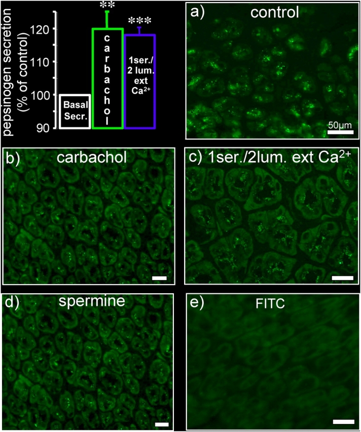
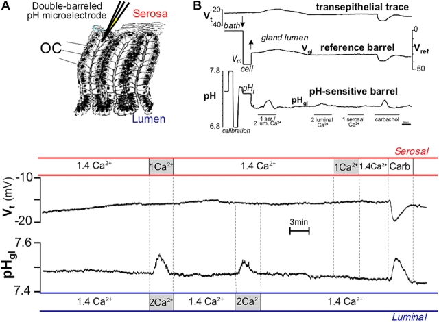
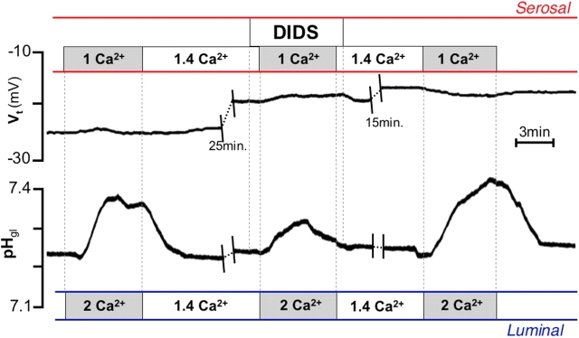
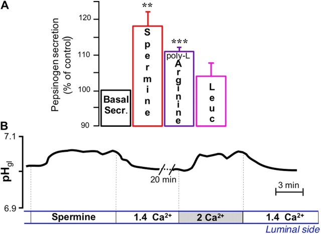
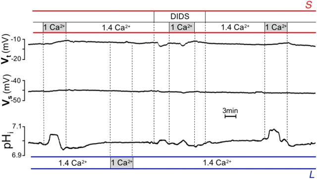
It is generally assumed that the functional consequences of stimulation with Ca2+ -mobilizing agonists are derived exclusively from the second messenger action of intracellular Ca2+, acting on targets inside the cells. However, during Ca2+ signaling events, Ca2+ moves in and out of the cell, causing changes not only in intracellular Ca2+, but also in local extracellular Ca2+. The fact that numerous cell types possess an extracellular Ca2+ "sensor" raises the question of whether these dynamic changes in external [Ca2+] may serve some sort of messenger function. We found that in intact gastric mucosa, the changes in extracellular [Ca2+] secondary to carbachol-induced increases in intracellular [Ca2+] were sufficient and necessary to elicit alkaline secretion and pepsinogen secretion, independent of intracellular [Ca2+] changes. These findings suggest that extracellular Ca2+ can act as a "third messenger" via Ca2+ sensor(s) to regulate specific subsets of tissue function previously assumed to be under the direct control of intracellular Ca2+.


Figures

Figure 1.
Figure 1.
Pepsinogen secretion: effect of carbachol, bilateral changes in [Ca 2+ ] ext , and spermine. Top left: effect of 100 μM serosal carbachol (n = 5) and simultaneous luminal increase (from 1.4 to 2.0 mM) and serosal decrease (from 1.4 to 1.0 mM) in [Ca2+]ext (n = 6) on pepsinogen secretion in the stomach of Rana esculenta. These, as well as all other experiments described in the paper, were performed in the presence of the histamine H2 receptor blocker cimetidine to prevent acid secretion (serosal, 100 μM). Peak pepsinogen release was calculated as percentage of total pepsin activity. All data are expressed as mean ± SEM. **, P < 0.01; ***, P < 0.001. (a–e) Immunofluorescence of pepsinogen in transverse sections of frog stomach. Sections were treated with an affinity-purified mAb against human pepsinogen I and with fluorescein-conjugated goat anti–mouse IgG. (a) Positive pepsinogen staining in control mucosa; the enzyme appears packed in granules abundantly distributed in the cytoplasm. (b–d) Effect of exposure to carbachol, bilateral changes in [Ca2+]ext, or serosal spermine (1 mM). (e) Secondary antibody alone. Bars, 50 μm.
Figure 2.
Figure 2.
Alkaline secretion: effect of carbachol and bilateral changes in [Ca 2+ ] ext . (A) Inset: schematic drawing of the technique used to record pH (or Ca2+) in the gland lumen. Double-barreled pH or Ca2+-sensitive microelectrodes were inserted in the gland lumen of a single gastric gland of the intact amphibian gastric mucosa perfused in vitro. (B) Top portion: original trace recording showing the entire experimental protocol. Top trace: transepithelial potential (Vt), luminal surface negative; middle trace: cell membrane potential (Vm) and gland lumen potential (Vgl) as measured by the reference barrel; bottom trace: cell pH (pHi) and pH in the gland lumen (pHgl) as measured by the pH-selective barrel. The microelectrode was calibrated before the puncture by flushing the chamber with Hepes-buffered Ringer's solutions with pH between 6.8 and 7.8. Afterward, the microelectrode was first inserted into a cell (arrow down, Vm and pHi) and then was further advanced into the gland lumen (arrow up) where the potential recording suddenly changed to nearly the same values as those measured transepithelially (Vgl). A similar experiment with longer baselines before and after responses is depicted in the bottom portion in expanded format (as used in all subsequent figures of this paper), with the reference barrel recording (used to ascertain proper positioning of the tip; Debellis et al., 1998) omitted for simplicity. Top bars (red) show changes in the serosal and bottom bars (blue) in the luminal solution. Vt did not change in response to [Ca2+]ext alterations, but it increased after 100 μM carbachol (by −6.3 ± SEM 0.9 mV; P < 0.0001, n = 10); pHgl increased by 0.13 ± 0.01 (P < 0.0001) in response to bilateral changes in [Ca2+]ext, by 0.06 ± 0.01 (P < 0.0001) in response to unilateral increase in luminal [Ca2+]ext, and by 0.11 ± 0.01 pH units (P < 0.0001) in response to carbachol, but did not change in response to unilateral decrease in serosal [Ca2+]ext (n = 10).
Figure 3.
Figure 3.
Effect of DIDS on alkaline secretion elicited by bilateral [Ca2 + ]ext changes. Exposure to 200 μM serosal DIDS resulted in significant reduction of the response to [Ca2+]ext changes (from 0.10 ± 0.02 to 0.04 ± 0.01 pH units; P < 0.01, n = 7).
Figure 4.
Figure 4.
Involvement of CaR: effect of spermine and amino acids. (A) Effect of 1 mM luminal spermine (n = 8), 1 mM poly-l-arginine (n = 3), and 1 mM leucine (n = 3) on basal pepsinogen secretion. **, P < 0.02; ***, P < 0.001. (B) Effect of luminal spermine and of increase in luminal [Ca2+]ext on pHgl. The increase in pHgl elicited by spermine (0.06 ± 0.01 pH units; P < 0.01, n = 7) was similar to that elicited by increase in luminal [Ca2+]ext. Spermine and high luminal [Ca2+]ext did not affect Vt (not depicted).
Figure 5.
Figure 5.
Effect of buffering luminal [Ca 2+ ] ex t increase. (A) Measurement of [Ca2+]ext in the gland lumen with Ca2+-sensitive double-barreled microelectrodes. Inset: Ca2+ buffering power of citrate Ringer's solution. Graph shows effect on potential measured by Ca2+-selective microelectrode (−VCa 2+) on addition of 200-μM aliquots of CaCl2 (arrows). Starting solution contained 1.4 mM added CaCl2; buffering power of citrate solution was at least twice as great as that of control solution at 2 mM Ca2+. Carbachol was not able to elicit an increase in luminal [Ca2+]ext in the presence of the buffer (348.0 ± 70.6 before vs. 23.7 ± 24.0 μM after addition of citrate; P < 0.01, n = 4). (B) Top: in the presence of citrate buffer, basal pepsinogen secretion did not change significantly after carbachol (n = 4), but did increase significantly in response to spermine (*, P < 0.05; n = 5). Bottom: in the presence of citrate buffer, carbachol still increased Vt (by −6.7 ± 1.34, top trace), but was no longer able to stimulate alkaline secretion (by −0.01 ± 0.01, bottom trace). In the same gland, spermine, in the presence of carbachol and citrate, increased pHgl (by 0.060 ± 0.001 pH units; P < 0.01, n = 4). The pHgl response to carbachol was recovered after removal of the buffer.
Figure 6.
Figure 6.
Responses are independent of intracellular Ca 2+ signals. (A) After prolonged (20 min) exposure to the sarco-ER ATPase pump inhibitor tBHQ (15 μM), spermine was still able to stimulate the increase in pHgl (0.070 ± 0.001 pH units before and 0.08 ± 0.01 after tBHQ; n = 4). (B) After pretreatment with 50 μM BAPTA-AM, spermine was still able to alkalinize the gland lumen. In the same glands, the response to carbachol was instead significantly depressed by BAPTA-AM (top, n = 3). ***, P < 0.002.
Figure 7.
Figure 7.
Intracellular Ca 2+ does not change after stimulation with spermine or external Ca 2+ . Changes in OC [Ca2+]i recorded with intracellular double-barreled Ca2+-sensitive microelectrodes. Potentials recorded by the selective barrel are displayed as VCa and have not been converted to free [Ca2+]i; a decrease in potential indicates an increase in [Ca2+]i. Only traces from the selective barrel are shown. Top: response to 2 mM luminal spermine and 100 μM carbachol. Middle: effect of bilateral changes in [Ca2+]ext and 10 μM ionomycin. Bottom: summary of responses. **, P < 0.01; ***, P < 0.001.
Figure 8.
Figure 8.
Involvement of the cAMP pathway. (A) The AC inhibitor SQ 22,536 (100 μM) increased pH in the gland lumen, mimicking the response elicited by spermine in the same glands. The responses to SQ 22,536 and spermine were comparable (top, n = 3). Spermine added on top of SQ 22,536 did not elicit any additional alkalinization of the gland lumen (ΔpH = −0.010 ± 0.006 pH units; n = 4). (B) Pretreatment with the Gi inhibitor pertussis toxin (PTX) abolished the increase in pHgl normally elicited by spermine (ΔpH = 0.085 ± 0.006 pH units before and 0.013 ± 0.008 after PTX; n = 4; **, P < 0.01) or carbachol (ΔpH = 0.087 ± 0.003 pH units before and 0.010 ± 0.010 after PTX; n = 3; **, P < 0.01). However, SQ 22,536 was still able to evoke gland lumen alkalinization after PTX treatment (ΔpH = 0.093 ± 0.020 pH units before and 0.093 ± 0.019 after PTX; n = 3). Vt response to carbachol stimulation was not affected by PTX. Top: summary of responses to 2 mM luminal spermine, 100 μM carbachol, and 100 μM SQ 22,536 after a 1-h treatment with 0.25 μg/ml PTX. Left: effect on alkaline secretion; right: effect on Vt. Bottom: trace recording showing responses to luminal spermine, carbachol, and SQ 22,536 after treatment with PTX.
Figure 9.
Figure 9.
Effect of lowering serosal [Ca2 + ] on intracellular pH. Decreasing serosal Ca2+ led to an increase in cell pH (pHi) that was reversibly inhibited by serosal application of 200 μM DIDS (from 0.13 ± 0.01 to 0.04 ± 0.02 pH units; P < 0.01, n = 6). This maneuver did not change transepithelial or serosal membrane potentials (Vt and Vs, respectively). High luminal Ca2+did not affect pHi, Vs, or Vt.

Similar articles

Cited by

References

    1. Ashby, M.C., and A.V. Tepikin. 2002. Polarized calcium and calmodulin signaling in secretory epithelia. Physiol. Rev. 82:701–734. - PubMed
    1. Babini, E., M. Paukert, H.S. Geisler, and S. Grunder. 2002. Alternative splicing and interaction with di- and polyvalent cations control the dynamic range of acid-sensing ion channel 1 (ASIC1). J. Biol. Chem. 277:41597–41603. - PubMed
    1. Belan, P., O. Gerasimenko, A. Tepikin, and O. Petersen. 1996. Localization of Ca2+ extrusion sites in pancreatic acinar cells. J. Biol. Chem. 271:7615–7619. - PubMed
    1. Belan, P., O. Gerasimenko, O.H. Petersen, and A.V. Tepikin. 1997. Distribution of Ca2+ extrusion sites on the mouse pancreatic acinar cell surface. Cell Calcium. 22:5–10. - PubMed
    1. Berridge, M.J., M.D. Bootman, and H.L. Roderick. 2003. Calcium signalling: dynamics, homeostasis and remodelling. Nat. Rev. Mol. Cell Biol. 4:517–529. - PubMed

Publication types