. 2004 Jul 15;173(2):1033-42.
doi: 10.4049/jimmunol.173.2.1033.

Specific engagement of TLR4 or TLR3 does not lead to IFN-beta-mediated innate signal amplification and STAT1 phosphorylation in resident murine alveolar macrophages

Affiliations

Specific engagement of TLR4 or TLR3 does not lead to IFN-beta-mediated innate signal amplification and STAT1 phosphorylation in resident murine alveolar macrophages

Antonello Punturieri et al. J Immunol. .

Abstract

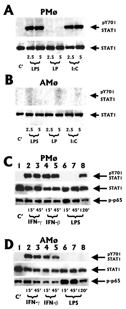
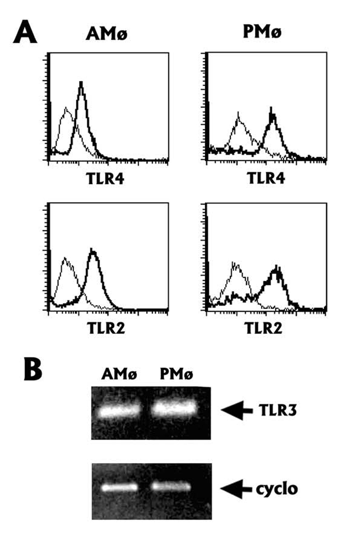
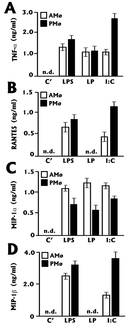
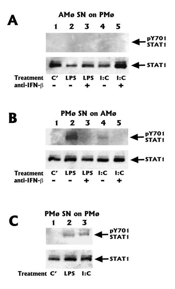
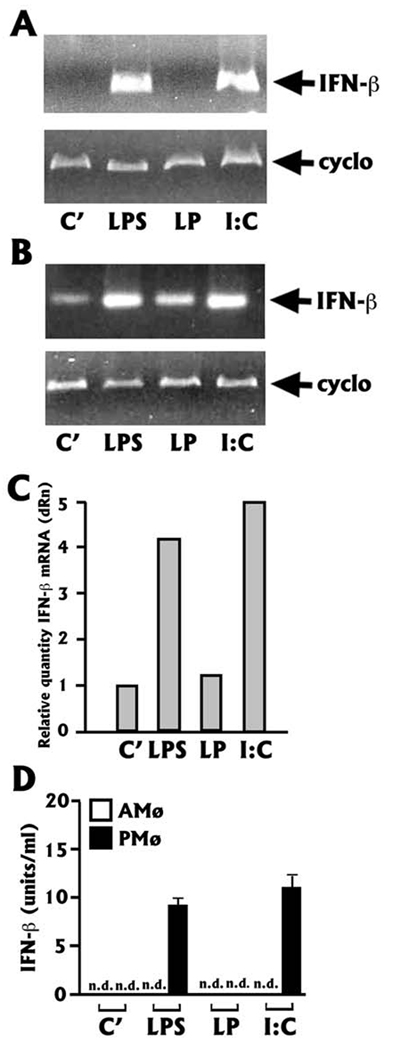
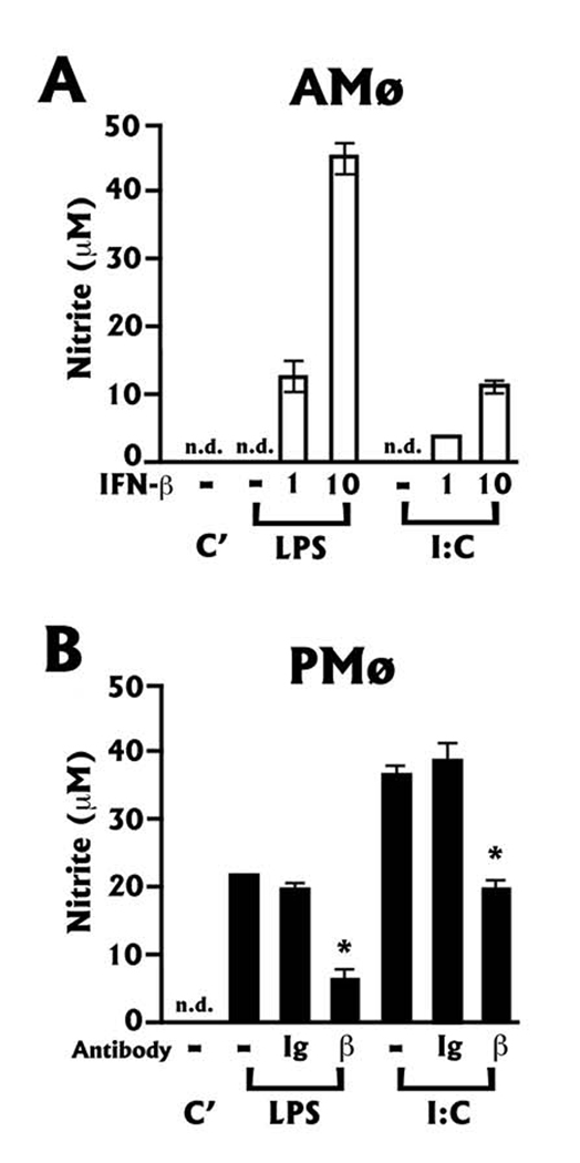
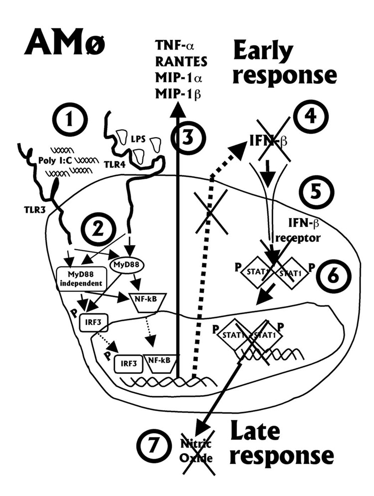
The innate immune response must be mobilized promptly yet judiciously via TLRs to protect the lungs against pathogens. Stimulation of murine peritoneal macrophage (PMphi) TLR4 or TLR3 by pathogen-associated molecular patterns (PAMPs) typically induces type I IFN-beta, leading to autocrine activation of the transcription factor STAT1. Because it is unknown whether STAT1 plays a similar role in the lungs, we studied the response of resident alveolar macrophages (AMphi) or control PMphi from normal C57BL/6 mice to stimulation by PAMPs derived from viruses (polyriboinosinic:polyribocytidylic acid, specific for TLR3) or bacteria (Pam(3)Cys, specific for TLR2, and repurified LPS, specific for TLR4). AMphi did not activate STAT1 by tyrosine phosphorylation on Y701 following stimulation of any of these three TLRs, but readily did so in response to exogenous IFN-beta. This unique AMphi response was not due to altered TLR expression, or defective immediate-early gene response, as measured by expression of TNF-alpha and three beta chemokines. Instead, AMphi differed from PMphi in not producing bioactive IFN-beta, as confirmed by ELISA and by the failure of supernatants from TLR-stimulated AMphi to induce STAT1 phosphorylation in PMphi. Consequently, AMphi did not produce the microbicidal effector molecule NO following TLR4 or TLR3 stimulation unless exogenous IFN-beta was also added. Thus, murine AMphi respond to bacterial or viral PAMPs by producing inflammatory cytokines and chemokines, but because they lack the feed-forward amplification typically mediated by autocrine IFN-beta secretion and STAT1 activation, require exogenous IFN to mount a second phase of host defense.


Figures

FIGURE 1
FIGURE 1
Murine AMø do not phosphorylate STAT1 in response to TLR engagement, but do so in response to exogenous IFNs. A, B. TLR engagement. Resident PMø (panels A) or AMø (panels B) were incubated for 2.5 or 5h in the presence of medium alone (lane C’), 100 ng/ml of LPS (lanes LPS), 100 ng/ml of LP (lanes LP) or 50 µg/ml of poly I:C (lanes I:C). Cells were then harvested and processed for Western blot analysis as specified in the Materials and Methods section. The same membranes were first probed with anti-pY701 STAT1 (panel A, B top), and then were stripped and re-probed with anti-STAT1 (panel A, B bottom). pY701 STAT1, anti-phospho STAT1 antibody that recognizes the tyrosine 701 phosphorylated forms of both p92 and p84 STAT1. STAT1, anti-STAT1 antibody that recognizes both p92 and p84 STAT1. One of three independent experiments with identical results is shown. C, D. Effect of exogenous IFNs. Resident PMø (panels C) or AMø (panels D) were stimulated for the indicated times with 100 U/ml of IFN-γ (lanes IFN-γ), 10,000 U/ml of IFN-β (lanes IFN-β) or 100 ng/ml of LPS (lanes LPS). The same membranes were first probed with anti-pY701 STAT1 (panels C, D top), and then were stripped and re-probed with anti-STAT1 (panels C, D middle) or with anti-phospho p65 NF-kB subunit (panels C, D bottom). One of two independent experiments with identical results is shown.
FIGURE 2
FIGURE 2
Murine AMø express TLR4, TLR2 and TLR3 receptors. A. Flow cytometric analysis of TLR4/MD-2 (panel A, top) or TLR2 (panel A, bottom) receptors expression in AMø (A, left panels) and PMø (A, right panels). The heavy line profile shows specific staining, the light line profile shows staining with isotype-matched control IgGs. Representative histograms are shown from two independent experiments with nearly identical results. B. RT-PCR analysis of TLR3 expression. Un-stimulated AMø (B, left lane) or PMø (B, right lane) mRNA was harvested and analyzed for TLR3 expression by RT-PCR as specified in the Materials and Methods section. Cyclo is the housekeeping gene cyclophilin, which was used to adjust the volume of the cDNA input used in each reaction to reach apparent equal expression of the final PCR products. One of two independent experiments with nearly identical results is shown.
FIGURE 3
FIGURE 3
Production of TNF-α and beta chemokines indicates that both the MyD88-dependent and the MyD88-independent pathways are functional in resident AMø. Resident AMø (open bars) and PMø (closed bars) from normal C57BL/6 mice were cultured in the presence of the indicated stimuli for 6h, the supernatant collected and processed for ELISA for TNF-α (A); RANTES (B); MIP-1α (C); and MIP-1β (D). Mø were stimulated in the presence of medium alone (lane C’), 100 ng/ml of LPS (lane LPS), 100 ng/ml of LP (lane LP) or 50 µg/ml of poly I:C (lane I:C). n.d., none detected. Note the difference in scales between the panels. Data are mean ± SEM of triplicate wells in each of four independent experiments.
FIGURE 4
FIGURE 4
AMø do not release bioactive IFN-β in response to TLR4 or TLR3 stimulation. Resident PMø (panels A, C) or AMø (panel B) (responding cells) were exposed to supernatant (SN) from resident AMø (panel A) or to supernatant from PMø (panels B, C) which had been stimulated for 3 h with medium alone (lane C’), 100 ng/ml LPS (lanes LPS) or 50 µg/ml poly I:C (lanes I:C). Before addition to the responding cells, all supernatant were treated with 10 µg/ml of polymixin B. Some supernatant were also incubated with 5,000 neutralization units of anti-IFN-β antibody for 30 min (panels A–B, lanes 3 and 5). After 30 min, adherent responder Mø were washed, lysed, and assayed by Western blotting. The same membrane was first probed with anti-pY701 STAT1 (panels A–C top), and then stripped and re-probed with anti-STAT1 (panels A–C bottom). One of two independent experiments with identical results is shown.
FIGURE 5
FIGURE 5
Regulation of IFN-β mRNA and protein in AMø upon TLR stimulation. A, B. RT-PCR mRNA analysis. Resident PMø (panels A) or AMø (panels B) were incubated for 2h in the presence of medium alone (lane C’), 100 ng/ml of LPS (lane LPS), 100 ng/ml LP (lane LP) or 50 µg/ml poly I:C (lane I:C). Mø mRNA was harvested and analyzed by RT-PCR as specified in the Materials and Methods section. Cyclo is the housekeeping gene cyclophilin, which was used to adjust the volume of the cDNA input used in each reaction to reach apparent equal expression of the final PCR products. One of three independent experiments with nearly identical results is shown. C. Real-Time RT-PCR. Total RNA was extracted from AMø treated as specified above and analyzed for IFN-β mRNA expression as described in the Materials and Methods section. The relative quantities of specific mRNA from treated AMø are plotted compared to the calibrator mRNA (IFN-β mRNA in control AMø defined as 1). dRn is baseline corrected, reference dye-normalized fluorescence. Since replicates are treated collectively eliminating replicate variability, no standard deviation or coefficient of variation are showed. D. IFN-β ELISA. Supernatants from resident AMø (open bars) and PMø (closed bars) from normal C57BL/6 mice cultured in the presence of the indicated stimuli for 3h were analyzed by a custom-made ELISA assay. Mø were stimulated in the presence of medium alone (lane C’), 100 ng/ml of LPS (lane LPS), 100 ng/ml of LP (lane LP) or 50 µg/ml of poly I:C (lane I:C). n.d., none detected. Data are mean ± SEM of triplicate wells in each of two independent experiments.
FIGURE 6
FIGURE 6
Nitrite accumulation in the supernatant of AMø and PMø after exposure to TLR4 or TLR3 stimulants depends on IFN-β. Resident AMø (A) or PMø (B) were stimulated for 72 h with medium alone (lane C’), 100 ng/ml of LPS (lanes LPS) or 50 µg/ml of poly I:C (lanes I:C). In panel A, resident AMø were also incubated with 1,000 (1) or 10,000 (10) units/ml of recombinant IFN-β. B, resident PMø were incubated with either control rabbit immunoglobulins (Ig) or 8,000 neutralizing units/ml of anti-IFN-β Ab (β) at the beginning of the experiment. Supernatants from these cultures were assayed for the accumulation of nitrite using the Griess reagent as described in the Materials and Methods section. Neither 1,000 nor 10,000 units/ml of IFN-β alone resulted in NO production by AMø or PMø (not shown). One of two independent experiments performed in triplicate is shown. *, p<0.05, Student t-test.
FIGURE 7
FIGURE 7
Schematic model of the differences between AMø and PMø in response to TLR4 or TLR3 stimulation. In both resident AMø and PMø. (1) activation of TLR4 or TLR3 by bacterial- or viral-derived PAMPs leads (2) via the MyD88-dependent and MyD88-independent pathways to activation of IRF3 and NF-κB. For clarity, the other IFN-β essential transcription factors ATF-2 and c-Jun are omitted. These transcription factors induce elaboration of immediate early genes (3) typified by TNF-α, RANTES, MIP-1α and MIP-1β by both types of Mø. AMø, however, do not go on to produce (4) and/or secrete IFN-β. Consequently, (5, 6) STAT1 is not activated and there is defective elaboration of the second cascade of anti-microbial mediators, (7) typified by NO.

Similar articles

Cited by

References

    1. Barnes PJ. New concepts in chronic obstructive pulmonary disease. Annu Rev Med. 2003;54:113. - PubMed
    1. Tetley TD. Macrophages and the pathogenesis of COPD. Chest. 2002;121:156S. - PubMed
    1. Newman SL, Henson JE, Henson PM. Phagocytosis of senescent neutrophils by human monocyte-derived macrophages and rabbit inflammatory macrophages. J Exp Med. 1982;156:430. - PMC - PubMed
    1. Hu B, Sonstein J, Christensen PJ, Punturieri A, Curtis JL. Deficient in vitro and in vivo phagocytosis of apoptotic T cells by resident murine alveolar macrophages. J Immunol. 2000;165:2124. - PMC - PubMed
    1. Hodge S, Hodge G, Scicchitano R, Reynolds PN, Holmes M. Alveolar macrophages from subjects with chronic obstructive pulmonary disease are deficient in their ability to phagocytose apoptotic airway epithelial cells. Immunol Cell Biol. 2003;81:289. - PubMed

Publication types

MeSH terms