Comparative Study
. 2005 Jan;93(1):267-80.
doi: 10.1152/jn.00747.2003. Epub 2004 Jul 7.

Comparative morphology of rodent vestibular periphery. II. Cristae ampullares

Collaborators, Affiliations
Comparative Study

Comparative morphology of rodent vestibular periphery. II. Cristae ampullares

Sapan S Desai et al. J Neurophysiol. 2005 Jan.

Erratum in

Abstract

We made flattened neuroepithelial preparations of horizontal and vertical (anterior and posterior) cristae from mouse, rat, gerbil, guinea pig, chinchilla, and tree squirrel. Calretinin immunohistochemistry was used to label the calyx class of afferents. Because these afferents are restricted to the central zone of the crista, their distribution allowed us to delineate this zone. In addition to calyx afferents, calretinin also labels approximately 5% of type I hair cells and 20% of type II hair cells throughout the mouse and rat crista epithelium. Measurements of the dimensions of the cristae and counts of hair cells and calyx afferents were determined on all species. Numbers of calyx afferents, hair cells, area, length, and width of the sensory epithelium increase from mouse to tree squirrel. As in the companion paper, we obtained additional data on vestibular end organ dimensions from the literature to construct a power law function describing the relationship between crista surface area and body weight. The vertical cristae of the mouse, rat, and gerbil have an eminentia cruciatum, a region located transversely along the midpoint of the sensory organ and consisting of nonsensory cells. Apart from this eminentia cruciatum, there are no statistical differences between horizontal and vertical cristae with regard to area, width, length, the number and type of hair cells, and number of calretinin-labeled calyx afferents.


Figures
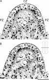
FIG. 1
FIG. 1
The use of the disector method for hair cell counts. These are consecutive 2-μm sections through the transverse axis of a chinchilla horizontal crista. Adjacent 2-μm sections every 50 μm were used in each species to estimate the population of type I, type II, and supporting cells (small arrows). A: reference section; B: lookup section. Nuclei from hair cells located in A but not in B are coded in white; nuclei from hair cells located in both sections are coded in black. A similar procedure was done for supporting cell counts (not shown for clarity). The reference section and lookup section were then reversed and the process was repeated to provide a 2nd independent set of counts. Averaging the counts from these 2 sections yielded Q. Cells were distinguished by type, and the numbers were extrapolated to estimate the population. The plane of section is shown in the inset. Scale bar =50 μm.
FIG. 2
FIG. 2
A standard flattened reconstruction of a chinchilla crista with our 2 zones defined. The central zone (CZ), shaded in light gray, is the inner zone. The outer zone is the peripheral zone (PZ). The area of the central zone (ACZ) and area of the sensory epithelium (ASE, contained within the outermost lines) were measured. The length of the central zone (LCZ) and length of the crista (LCR) were measured. The width of the central zone (WCZ) is also indicated. The width of the crista was determined at the midpoint (WM), which is also the narrowest point of the crista. The width of the crista at the planum (WP), the widest point, was determined for both ends and averaged. Mean values are presented in Table 1. Scale bar = 100 μm.
FIG. 3
FIG. 3
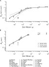
Area of crista related to body weight and to the area of the saccular and utricular maculae. A: relationship between body weight and the area of the sensory epithelium (ASE) in cristae (closed symbols). Dashed line extends the analysis in the present study to data taken from other species in the literature (open symbols, see figure). B: the ratios of the sensory epithelium area for these 3 sensory organs were compared. The species used in this panel (mouse, rat, gerbil, guinea pig, chinchilla, squirrel monkey, tree squirrel, hare, cat, human, pig, cow) can be distinguished by aligning the body weights with those in A. All 3 fits of surface area vs. body weight were normalized to an average of the 3 fits. A linear correlation between crista sensory epithelium area (open squares) and that of the saccular macula (open diamonds) and utricular macula (open circles) was performed. Note that the utricular macula is disproportionately larger for species >5kg in weight.
FIG. 4
FIG. 4
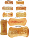
Horizontal and vertical cristae from the 6 species used in this study are shown. Mouse, rat, and gerbil superior cristae (SC) and posterior cristae (PC) have a nonsensory region of supporting cells near the midpoint of the sensory organ, known as the eminentia cruciatum. This region is not found in the vertical cristae of guinea pig, chinchilla, or tree squirrel nor in the horizontal cristae (HC) of any of the species examined in this study. The horizontal cristae of guinea pig, chinchilla, and tree squirrel appear similar to their vertical cristae counterparts, and are not shown. Scale bar = 100 μm.
FIG. 5
FIG. 5
Schematic of cristae with zonal boundaries drawn and calyx terminals coded by the number of type I hair cells they contact. The central zone is delineated by a thin black line, and the extent of the sensory epithelium is outlined by a thicker black line. The calretinin-labeled calyx afferents are coded by colored dots indicating the complexity of the afferent. Simple calyx afferents (red) contact a single hair cell. Double complex calyx afferents (green) contact 2 hair cells. Triple complex calyx afferents (blue) contact 3 hair cells. Higher levels of complex calyx afferents (yellow), found in the guinea pig, chinchilla, and tree squirrel, may contact ≤5 type I hair cells (see Table 4). Scale bar = 100 μm.
FIG. 6
FIG. 6
A Western blot to confirm the molecular weight of calretinin and the specificity of our antibody. The Western blot allowed us to determine that the staining in the mouse and rat type I and type II hair cells was calretinin. For mouse (Ms), rat (Rt), and chinchilla (Ch) cristae and vestibular ganglia (VG), only 1 band was found, corresponding to the 29-kDa molecular weight (MW) of calretinin. Our positive control, rat cerebellum (Cblm), also had a similar band. We used rat kidney (kidney) as a negative control.
FIG. 7
FIG. 7
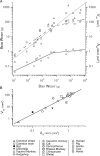
Relations among vestibular structures, brain weight, and body weight. A: body weight (BW, abscissa) is plotted vs. surface area of the crista sensory epithelium (ASEcrista, right axis), vestibular nuclear volume (VVN, right axis), and brain weight (left axis). Values for individual species points and linear fits are coded, respectively, as follows: ASEcrista (○ — ○), VVN (□ - - - □), and BW (△ • • • △). For the animals in this series, the brain grows as a power law with a slope of 0.78. In contrast, both the ASEcrista and the VVN grow with brain weight for small animals, but then the rate slows considerably for large animals (>4 kg). B: crista surface area (ASEcrista) and vestibular nuclear volume (VVN) are plotted against 1 another (r2 = 0.95). VVN grows faster than ASEcrista, implying that as vestibular nuclear volume increases, each vestibular afferent has a more extensive set of central terminations and covers a larger volume of the vestibular nuclei. Values used for Fig. 7 were taken from both this study and the literature (coded symbols, see figure and see text for references).

References

    1. Baird RA, Desmadryl G, Fernández C, Goldberg JM. The vestibular nerve of the chinchilla. II. Relation between afferent response properties and peripheral innervation patterns in the semicircular canals. J Neurophysiol. 1988;60:182–203. - PubMed
    1. Bäurle J, Guldin W. Unbiased number of vestibular ganglion neurons in the mouse. Neurosci Lett. 1998;246:89–92. - PubMed
    1. Blinkov SM, Ponomarev VS. Quantitative determinations of neurons and glial cells in the nuclei of the facial and vestibular nerves in man, monkey and dog. J Comp Neurol. 1965;125:295–302. - PubMed
    1. Boord RL, Rasmussen GL. Analysis of myelinated fibers of the acoustic nerve of the chinchilla (Abstract). Anat Rec. 1958;127:394.
    1. Brichta AM, Peterson EH. Functional architecture of vestibular primary afferents from the posterior semicircular canal of a turtle, Pseudemys (Trachemys) scripta elegans. J Comp Neurol. 1994;344:481–507. - PubMed

Publication types

MeSH terms

LinkOut - more resources