. 2004 Jul 7;24(27):6086-97.
doi: 10.1523/JNEUROSCI.5635-03.2004.

Redistribution of GABAB(1) protein and atypical GABAB responses in GABAB(2)-deficient mice

Affiliations

Redistribution of GABAB(1) protein and atypical GABAB responses in GABAB(2)-deficient mice

Martin Gassmann et al. J Neurosci. .

Abstract

GABAB receptors mediate slow synaptic inhibition in the nervous system. In transfected cells, functional GABAB receptors are usually only observed after coexpression of GABAB(1) and GABAB(2) subunits, which established the concept of heteromerization for G-protein-coupled receptors. In the heteromeric receptor, GABAB(1) is responsible for binding of GABA, whereas GABAB(2) is necessary for surface trafficking and G-protein coupling. Consistent with these in vitro observations, the GABAB(1) subunit is also essential for all GABAB signaling in vivo. Mice lacking the GABAB(1) subunit do not exhibit detectable electrophysiological, biochemical, or behavioral responses to GABAB agonists. However, GABAB(1) exhibits a broader cellular expression pattern than GABAB(2), suggesting that GABAB(1) could be functional in the absence of GABAB(2). We now generated GABAB(2)-deficient mice to analyze whether GABAB(1) has the potential to signal without GABAB(2) in neurons. We show that GABAB(2)-/- mice suffer from spontaneous seizures, hyperalgesia, hyperlocomotor activity, and severe memory impairment, analogous to GABAB(1)-/- mice. This clearly demonstrates that the lack of heteromeric GABAB(1,2) receptors underlies these phenotypes. To our surprise and in contrast to GABAB(1)-/- mice, we still detect atypical electrophysiological GABAB responses in hippocampal slices of GABAB(2)-/- mice. Furthermore, in the absence of GABAB(2), the GABAB(1) protein relocates from distal neuronal sites to the soma and proximal dendrites. Our data suggest that association of GABAB(2) with GABAB(1) is essential for receptor localization in distal processes but is not absolutely necessary for signaling. It is therefore possible that functional GABAB receptors exist in neurons that naturally lack GABAB(2) subunits.


Figures
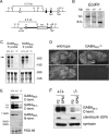
Figure 1.
Figure 1.
Characterization of GABAB(2)-/- mice. A, Top, GABAB(2) locus encompassing exons 8-11, encoding part of the N-terminal and the transmembrane (TM) domains 1 and 2. Bottom, GABAB(2) allele after homologous recombination with a targeting construct containing a neomycin resistance cassette (neo) flanked by 4.5 and 1.8 kb of genomic DNA (bold lines). Exons 9 and 10 (3.5 kb, black boxes) are deleted. The Southern blot probe used in B is indicated. H, HindIII; E, EcoRI; B, BamHI. B, Southern blot analysis of EcoRI cut genomic DNA from wild-type (+/+), GABAB(2)+/- (+/-), and GABAB(2)-/- (-/-) mice. C, Top, Northern blot analysis of total brain RNA hybridized with GABAB(2) cDNA probes upstream (5′ probe) and downstream of the deletion (3′ probe). The probes hybridize to a band just above the 28 S ribosomal RNA (arrow) in wild-type and GABAB(2)+/-, but not in GABAB(2)-/-, mice. Bottom, Blots from top panels stained with methylene blue, demonstrating equal loading of RNA. Ribosomal RNA bands (18 and 28 S) are labeled. D, In situ hybridization analysis of GABAB(1) (1, top) and GABAB(2) (2, bottom) transcripts of sagittal sections from adult wild-type and GABAB(2)-/- brains. E, Immunoblot analysis of brain extracts from adult mice using antibodies directed against C-terminal and N-terminal epitopes of GABAB(2) and GABAB(1). Antibodies to PSD-95 control for equal loading. GABAB(1a) (1a) and GABAB(1b) (1b) proteins are indicated. F, Immunoblot analysis demonstrating the presence of GABAB(1) protein in synaptic plasma membranes (SPM) purified from the P2 pellet (P2) of brain extracts of wild-type and GABAB(2)-/- mice. Antibodies to calreticulin show that the synaptic plasma membrane fraction is free of ER proteins. Equal loading of samples was controlled with anti-syntaxin antibodies. To detect putative truncated GABAB(2) proteins, we used 15% SDS-PAGE and N-terminal GABAB(2) antibodies. In all other immunoblot experiments, we used 10% SDS-PAGE.
Figure 2.
Figure 2.
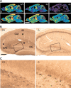
Alteration of GABAB receptor-IR in GABAB(2)-/- brains. A, Effect of GABAB(2) gene deletion on the distribution of GABAB(2)-IR (2) and GABAB(1)-IR (1), as visualized in color-coded parasagittal sections from adult wild-type (+/+), GABAB(2)+/- (+/-), and GABAB(2)-/- (-/-) mice. The color scale is indicated. The reduced expression of GABAB(2) in GABAB(2)+/- mice and the complete loss of expression in GABAB(2)-/- mice are evident throughout the brain (top). GABAB(1)-IR is retained in GABAB(2)+/- mice and partly reduced in GABAB(2)-/- mice, in which it exhibits an altered cellular distribution, as seen in the hippocampus (bottom). The residual GABAB(1)-IR in GABAB(2)-/- mice is not caused by nonspecific binding of the secondary antibodies, which are the same for GABAB(1) and GABAB(2). The specificity of the GABAB(1) antiserum was also tested in GABAB(1)-/- mice, in which no specific staining was observed (data not shown). B, Color photomicrographs of the hippocampal formation stained for GABAB(1) in adult wild-type and GABAB(2)-/- mice. The pronounced increase of IR in the CA1-CA3 pyramidal cell layer and in the dentate gyrus granule cell layer (DG) contrasts with the strong reduction in the dendritic layers [stratum oriens (so), stratum radiatum (sr), stratum lucidum (sl), and molecular layer (ml)]. C, Enlargement of the framed areas in B. Numerous interneurons, which are primarily hidden in sections from wild-type mice because of the homogeneous staining, appear more strongly labeled in GABAB(2)-/- mice but with a normal distribution and morphology. Scale bars: A, 2 mm; B, 200 μm.
Figure 6.
Figure 6.
Baclofen inhibits a postsynaptic K+ conductance in CA1 pyramidal cells of GABAB(2)-/- mice. A, Holding current (at -50 mV) plotted versus time for wild-type (top, filled circles) and GABAB(2)-/- (bottom, open circles) mice. Whereas both baclofen (50 μm) and adenosine (100 μm) induce an outward current in wild-type mice, baclofen induces an inward current in GABAB(2)-/- mice. Baclofen-induced effects were blocked by application of the GABAB receptor antagonist CGP55845A (2 μm) in wild-type as well as in GABAB(2)-/- mice. B, Summary graph illustrating the baclofen-induced inward current at -50 mV in GABAB(2)-/- mice. Baclofen-induced currents: wild-type, n = 5; GABAB(2)-/-, n = 9. Adenosine-induced currents: wild-type, n = 5; GABAB(2)-/-, n = 4. C, Current-voltage relationship of the baclofen-induced conductance in wild-type (black trace) and GABAB(2)-/- (gray trace) mice. Currents were obtained by calculating the difference between the I-V curves before and after addition of baclofen. Whereas a current with a positive slope conductance is induced by baclofen in wild-type mice, a current with negative slope conductance is induced in GABAB(2)-/- mice. D, Current-voltage relationship of the adenosine-induced conductance in wild-type mice (black trace) is not different from GABAB(2)-/- mice (gray trace). E, Baclofen induces the closure of K+ channels in GABAB(2)-/- mice. Raising extracellular [K+] concentration shifts the reversal potential of the baclofen-induced current. F, The baclofen-induced conductance change is mediated by G-protein activation. In the presence of intracellular GDPβS (1 mm for 25 min), both the baclofen-induced (control, n = 5; GDPβS, n = 5) and adenosine-induced (control, n = 4; GDPβS, n = 5) currents are inhibited in GABAB(2)-/- mice. G, Changes in the holding current (at -50 mV) in response to baclofen (Bacl.) after preincubation with adenosine. H, Summary graph illustrating that the effects of adenosine and baclofen are not fully additive. In wild-type neurons (+/+), the effect of a combined application of adenosine and baclofen is lower than the sum of the individual effects [Adenosine + Bacl. (calculated)]. In GABAB(2)-/- (-/-) neurons, the effects of adenosine and baclofen are not fully additive. Application of baclofen does not obliterate the adenosine response. I, Left, Summary graph illustrating postsynaptic conductance changes induced by baclofen in wild-type (n = 5), GABAB(2)+/- (n = 10), and GABAB(1)-/- (n = 4) mice. The conductance changes were blocked by application of the GABAB(1) receptor antagonist CGP55845A (2 μm; wild-type, n = 4; GABAB(2)-/-, n = 8). Adenosine-induced conductance changes are not different between genotypes (wild-type, n = 4; GABAB(2)-/-, n = 4; GABAB(1)-/-, n = 3). *p < 0.05; **p < 0.01.
Figure 3.
Figure 3.
GABAB(1) binding sites in GABAB(2)-/- brains. A, Saturation isotherms for [125I]CGP64213 antagonist binding to cortex membranes. No significant binding is detected in membranes from GABAB(2)-/- mice. The number of binding sites is reduced in GABAB(2)+/- versus wild-type mice. The maximal number of binding sites (Bmax) for wild-type and GABAB(2)+/- mice are 1.4 ± 0.12 and 0.7 ± 0.05 pmol/mg protein, respectively; Kd values were 1.1 ± 0.06 and 0.9 ± 0.05 nm, respectively (mean ± SEM; n = 3). B, Autoradiograms of brain extracts from wild-type (+/+), GABAB(2)+/- (+/-), and GABAB(2)-/- (-/-) mice, labeled with the photoaffinity antagonist [125I]CGP71872 (0.5 nm) and analyzed by SDS-PAGE. Exposure for 8 d (8d exp.) reveals low amounts of labeled GABAB(1a) (1a) and GABAB(1b) (1b) proteins in GABAB(2)-/- brains. C, GABAB(1) subunit autoradiography. Sagittal cryostat sections were incubated with the GABAB antagonist [3H]CGP62349. Nonspecific binding was determined in the presence of an excess of 100 μm unlabeled l-baclofen. Tritium-sensitive x-ray films were exposed for 24 hr and developed using a Cyclone Storage Phosphor screen (PerkinElmer Life Sciences, Boston, MA). D, Quantitative analysis of [3H]CGP62349 receptor autoradiography. Individual brain regions (n = 3) were counted using the MCID software package (Imaging Research, St. Catharines, Ontario, Canada). The differences in radioligand binding between the three genotypes are significant (two-sided Dunnett test; p < 0.001 for combined analysis of all brain regions). A-C, Representative experiments, which were repeated three times.
Figure 4.
Figure 4.
[35S]GTPγS binding to cortex membranes. No significant GABA-stimulated (filled symbols, filled lines) or baclofen-stimulated (Bac; open symbols, dotted lines) [35S] GTPγS binding is detected in GABAB(2)-/- membranes. [35S]GTPγS binding to membranes from GABAB(2)+/- mice is significantly reduced compared with wild-type mice. Values are normalized to the maximal response obtained with wild-type mice.
Figure 5.
Figure 5.
Lack of baclofen-induced presynaptic inhibition in CA1 pyramidal cells of GABAB(2)-/- mice. A, Excitatory synaptic transmission. Monosynaptic EPSC peak amplitudes plotted versus time and representative traces from wild-type (top, filled circles) and GABAB(2)-/- (bottom, open circles) mice. Both baclofen (50 μm) and adenosine (100 μm) significantly depress the EPSC amplitude in wild-type mice, whereas baclofen and CGP55845A (2 μm) have no effect on the EPSC amplitude in GABAB(2)-/- mice. The effect of adenosine is similar in both genotypes. Traces are averages of 10 consecutive sweeps. Calibration: 40 msec, 100 pA. B, Summary graph showing the lack of baclofen-induced presynaptic inhibition of excitatory synaptic transmission in GABAB(2)-/- mice (wild-type, n = 4; GABAB(2)-/-, n = 8). Adenosine-induced inhibition is similar in both genotypes (wild-type, n = 4; GABAB(2)-/-, n = 6). C, Inhibitory synaptic transmission. Monosynaptic IPSC peak amplitudes plotted versus time and representative traces from wild-type (top, filled circles) and GABAB(2)-/- (bottom, open circles) mice. Both baclofen (50 μm) and the μ-opioid agonist DAMGO (1 μm) significantly depress the IPSC amplitude in wild-type mice, whereas baclofen and CGP55845A (2 μm) have no effect on the IPSC amplitude in GABAB(2)-/- mice. The effect of DAMGO was similar in both genotypes. Traces are averages of 10 consecutive sweeps. Calibration: 100 msec, 200 pA. D, Summary graph showing the lack of baclofen-induced presynaptic inhibition of inhibitory synaptic transmission in GABAB(2)-/- mice (wild-type, n = 7; GABAB(2)-/-, n = 6). DAMGO-induced inhibition was similar in both genotypes (wild-type, n = 7; GABAB(2)-/-, n = 5). **p < 0.01; ***p < 0.001.
Figure 7.
Figure 7.
Lack of baclofen-induced delta waves in GABAB(2)-/- mice. A, Effect of l-baclofen (10 mg/kg, i.p.) on the EEG of freely moving wild-type (+/+) and GABAB(2)-/- (-/-) mice. The EEG of wild-type and GABAB(2)-/- mice were similar 10 min before baclofen application (-10 min). Twenty minutes after baclofen application, delta waves were observed in the EEG of wild-type, but not of GABAB(2)-/-, mice (+20 min). Single spikes appeared sporadically in the EEG of wild-type mice (+40 min), followed by delta waves that lasted for several hours (+7 hr). Ten hours after baclofen application, the EEG traces of wild-type and GABAB(2)-/- mice were again similar (+10 hr). B, Quantification of baclofen-induced delta waves in the EEG of wild-type and GABAB(2)-/- mice. The percentage of delta waves of the total power amplitude was calculated over periods of 10 min. Three to four mice per genotype were analyzed.
Figure 8.
Figure 8.
Lack of baclofen-induced motor impairment and hypothermia in GABAB(2)-/- mice. A, No baclofen-induced impairment of rotarod endurance is observed in GABAB(2)-/- (-/-) mice (n = 7-10). In contrast, wild-type mice (+/+) show a marked reduction in rotarod performance after baclofen application (p < 0.05; Fisher's post hoc tests). The vehicle-treated control groups stayed on the rotarod during the entire experiment (300 sec) at all time points examined. Thus, in the graph, the data points for the wild-type vehicle control (black dots) are hidden behind the data points for the GABAB(2)-/- vehicle control (white dots). At all time points after baclofen application (1, 2, and 4 hr), the GABAB(2)-/- group (white triangles) differed significantly from the wild-type control group (black triangles) (p < 0.05; Fisher's post hoc tests). All data points represent mean ± SEM values. B, Baclofen induces a potent reduction in body temperature in wild-type mice (black triangles) compared with the vehicle control group (black dots) (p < 0.05; Fisher's post hoc tests), whereas it is without effect on basal temperature in GABAB(2)-/- mice (n = 7-10). However, GABAB(2)-/- mice (white dots) exhibit a slight but significantly reduced basal temperature compared with wild-type littermates (black dots) (p < 0.05; Fisher's post hoc tests). All data points represent mean ± SEM values.
Figure 9.
Figure 9.
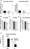
Behavioral analysis of GABAB(2)-/- mice. A, Hypolocomotor activity in GABAB(2)-/- mice. During a 1 hr observation period, GABAB(2) knock-out mice (-/-) moved over significantly longer distances (left histogram) with significantly greater speed (right histogram) than heterozygous (+/-) and wild-type (+/+) control mice. n = 7-8 per genotype; mean ± SEM; *p < 0.05. B, Response latencies of wild-type (+/+), heterozygous (+/-), and GABAB(2) knock-out (-/-) mice in the hotplate test assessed at 55°C. GABAB(2)-/- mice show significantly reduced paw-lick latencies compared with wild-type and heterozygous control groups. n = 19-20 per genotype; mean ± SEM; ***p < 0.001. C, Response latencies of wild-type (+/+), heterozygous (+/-), and GABAB(2) knock-out (-/-) mice in the tail-flick test assessed at infrared intensity 14. GABAB(2)-/- mice show significantly reduced tail-flick latencies compared with wild-type and heterozygous control groups. n = 19-21 per group; mean ± SEM; *p < 0.05. D, Paw-withdrawal thresholds for wild-type (+/+), heterozygous (+/-), and GABAB(2) knock-out (-/-) mice in response to a mechanical stimulus. Withdrawal thresholds of the left hindpaw were assessed for each genotype. GABAB(2)-/- mice show a significantly reduced withdrawal threshold compared with wild-type and heterozygous control groups. n = 19-21 per group; ***p < 0.001. Nociception tests were analyzed with Tukey's honestly significant difference test. In all tests, there were no significant differences in the behavior of wild-type or heterozygous mice. E, Impaired passive avoidance learning in GABAB(2)-/- mice. Step-through latencies of wild-type (+/+) and GABAB(2) knock-out (-/-) mice into the dark (shock) compartment on the training day (white bars) and in the retention test (black bars). GABAB(2)-/- mice were slower to enter on training day but faster in the retention test compared with the wild-type control mice. Wild-type, but not GABAB(2)-/-, mice show significantly longer latencies to enter the dark compartment in the retention test compared with the training trial, which is taken as an index of memory of the initial experience. n = 6-11 per group; mean ± SEM; ***p < 0.001 versus training; #p < 0.05 versus genotype; ##p < 0.01 versus genotype.

References

    1. Anderson WW, Collingridge GL (2001) The LTP Program: a data acquisition program for on-line analysis of long-term potentiation and other synaptic events. J Neurosci Methods 108: 71-83. - PubMed
    1. Bettler B, Kaupmann K, Mosbacher J, Gassmann M (2004) Molecular structure and physiological functions of GABAB receptors. Physiol Rev, in press. - PubMed
    1. Billinton A, Ige AO, Wise A, White JH, Disney GH, Marshall FH, Waldvogel HJ, Faull RL, Emson PC (2000) GABAB receptor heterodimer-component localisation in human brain. Brain Res Mol Brain Res 77: 111-124. - PubMed
    1. Blanchet C, Lüscher C (2002) Desensitization of mu-opioid receptor-evoked potassium currents: initiation at the receptor, expression at the effector. Proc Natl Acad Sci USA 99: 4674-4679. - PMC - PubMed
    1. Bouvier M (2001) Oligomerization of G-protein-coupled transmitter receptors. Nat Rev Neurosci 2: 274-286. - PubMed

Publication types

MeSH terms