. 2004 Jul 7;24(27):6218-27.
doi: 10.1523/JNEUROSCI.1692-04.2004.

Rapid axoglial signaling mediated by neuregulin and neurotrophic factors

Affiliations

Rapid axoglial signaling mediated by neuregulin and neurotrophic factors

Raymond M Esper et al. J Neurosci. .

Erratum in

  • J Neurosci. 2004 Jul 28;24(30):1 p following 6852

Abstract

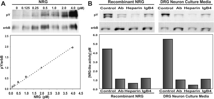
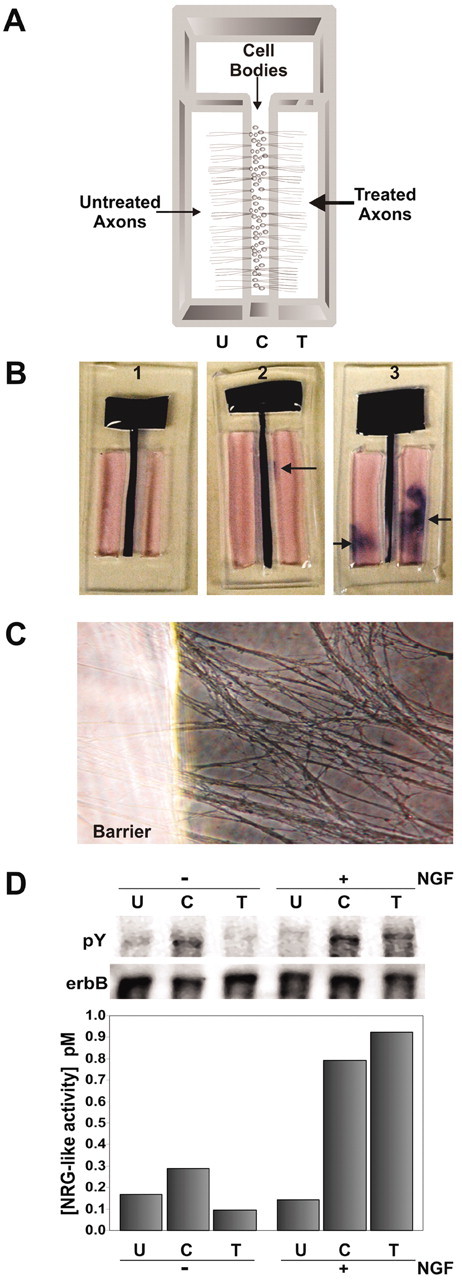
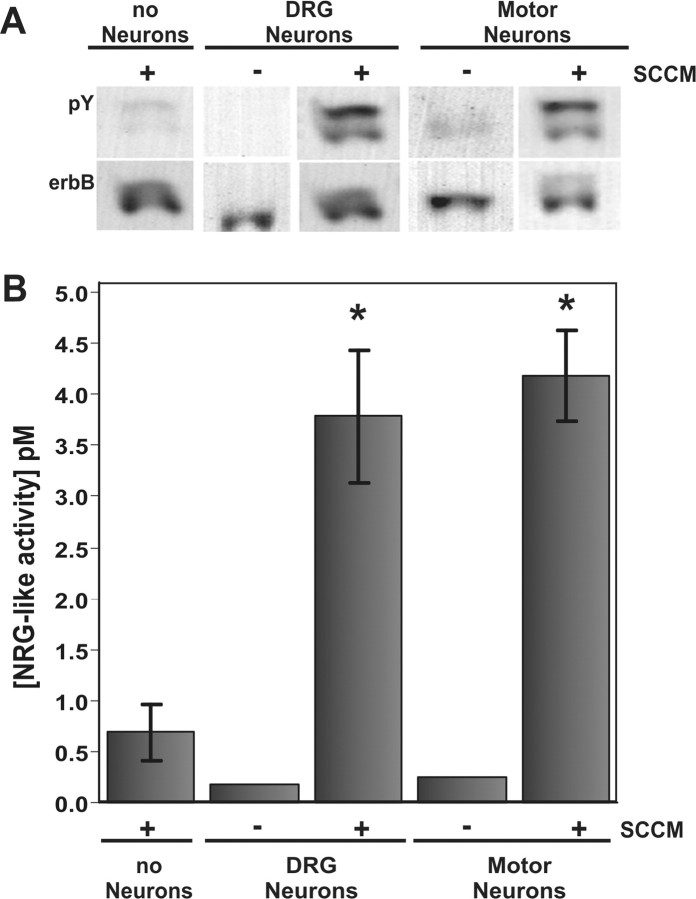
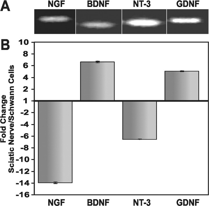
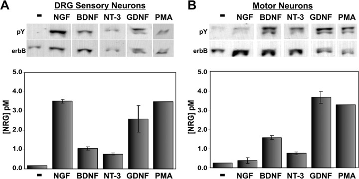
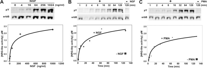
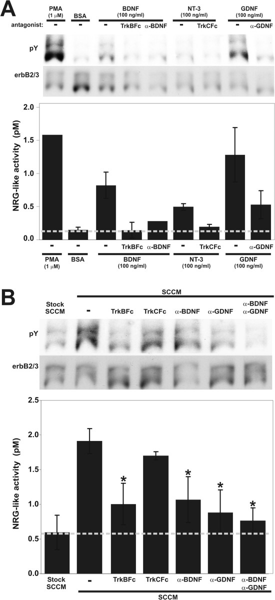
During peripheral nervous system development, Schwann cells are precisely matched to the axons that they support. This is mediated by axonal neuregulins that are essential for Schwann cell survival and differentiation. Here, we show that sensory and motor axons rapidly release heparin-binding forms of neuregulin in response to Schwann cell-derived neurotrophic factors in a dose-dependent manner. Neuregulin release occurs within minutes, is saturable, and occurs from axons that were isolated using a newly designed chamber slide apparatus. Although NGF and glial cell line-derived neurotrophic factor (GDNF) were the most potent neurotrophic factors to release neuregulin from sensory neurons, GDNF and BDNF were most potent for motor neurons and were the predominant neuregulin-releasing neurotrophic factors produced by cultured Schwann cells. Comparable levels of neuregulin could be released at a similar rate from neurons after protein kinase C activation with the phorbol ester, phorbol 12-myristate 13-acetate, which has also been shown to promote the cleavage and release of neuregulin from its transmembrane precursor. The rapid release of neuregulin from axons in response to Schwann cell-derived neurotrophic factors may be part of a spatially restricted system of communication at the axoglial interface important for proper peripheral nerve development, function, and repair.


Figures

Figure 1.
Figure 1.
Cultured neurons release minute quantities of soluble heparin-binding NRG. A, Western blot of immunoprecipitated erbB2 and erbB3 proteins from L6 myotubes treated with increasing amounts of NRG produces a sensitive and quantitative bioassay for NRG. The blot was probed with anti-phosphotyrosine (pY) antibodies and then stripped and reprobed with antibodies to erbB2 and erbB3 (erbB). Quantitation of this blot reveals the ratio of tyrosine-phosphorylated erbB protein to total erbB protein (pY/erbB) to be linear. The dashed line represents the linear regression of this data described by y = 0.4999x, R2 = 0.9936. B, Specificity of this NRG assay was shown using reagents that block the erbB receptor phosphorylation induced by recombinant NRG (5 pm) as well as sensory neuron culture media. Conditioned culture media was pretreated with an NRG-blocking antibody (1:10), soluble heparin (10 mg/ml), or a soluble fusion protein of erbB4 and human IgG (1:10) and then assayed for NRG as in A. Each reagent inhibited NRG-induced erbB tyrosine phosphorylation from the neuron cultures in a pattern identical to recombinant NRG, indicating that sensory neurons secrete predominately the soluble Ig form of NRG.
Figure 7.
Figure 7.
Focal release of NRG from axons. A, Schematic diagram of a compartmented Campenot-style chamber is shown where neurons were plated into the center compartment “C” and allowed to grow into both treated “T” and untreated “U” outer compartments. After 3d, the neurons extend axons equally into the outer compartments, whereas their cells bodies remain in the center. B, All culture chambers were tested individually for leakage between compartments at the end of each experiment by visual inspection of a tracker dye showing no leakage (1), minor leakage (2), or severe leakage (3). Only media from those chambers with no leakage were used for analysis. C, An example of axons growing under the clear silicone barrier (left) and into the outer compartment (right) is shown. D, These chamber slides were used to assess the effects of 100 ng/ml NGF for 2 hr on axons from the treated side compared with both the untreated and central compartments (right 3 lanes). Measurement of NRG in each of the three compartments revealed that distal axons in the NGF-treated compartment rapidly released NRG compared with the untreated side. NRG release was also promoted in the center compartment, containing proximal axons and cell bodies, suggesting a retrograde signaling event from the distal axons. Control culture chambers without NGF treatment (left 3 lanes) did not release as much NRG into either the T or C compartments. Media from a total of eight culture chambers was pooled for NGF treatment, and four culture chambers were pooled for the negative control. The same pattern of NRG release was seen in replicate experiments.
Figure 2.
Figure 2.
SCCM induces the rapid release of NRG from cultured neurons. A Western blot shown in A and then quantified in B showed that NRG release into both DRG sensory and motor neuron culture media was increased after exposure to SCCM for 2 hr. Although a small amount of NRG is present in the SCCM alone, primary sensory and motor neuron cultures released significantly more NRG in response to factors in the SCCM. This was performed in triplicate with the average and SD shown in the graph. Statistical significance for sensory (p = 0.0092) and motor (p = 0.0015) neurons was measured with a Student's t test.
Figure 3.
Figure 3.
Neurotrophic factors are expressed in sciatic nerve and Schwann cell cultures. A, NGF, BDNF, NT-3, and GDNF mRNAs can be detected in intact chicken E18 sciatic nerve using RT-PCR. B, Real-time, quantitative PCR showed dramatic differences in relative expression between intact sciatic nerve and 5-d-old Schwann cell cultures as follows: NGF (14-fold decreased), BDNF (6-fold increased), NT-3 (6-fold decreased), and GDNF (5-fold increased).
Figure 4.
Figure 4.
Neurotrophic factors induce NRG release from cultured sensory and motor neurons in distinct patterns. Western blots are shown for DRG sensory (A) and motor (B) neurons with quantitation of the amount of NRG released into neuronal culture media. Cultures were treated with either neurotrophic factors (100 ng/ml) or PMA (1 μm) for 2 hr. Although NGF and GDNF were most able to promote NRG release from sensory neurons, BDNF and GDNF were the two most potent factors for motor neurons. Activation of PKC with the phorbol ester PMA also induced maximal NRG release from both neuron types at a level comparable with neurotrophic factor treatment. Error bars correspond to 1 SD from three replicates.
Figure 5.
Figure 5.
Neurotrophin-induced NRG release from neurons is rapid and dose dependent. A, Primary sensory neurons were exposed to increasing concentrations of NGF for 2 hr. Released NRG was measured by the Western blot bioassay (top) with quantitation (bottom). NGF (4 ng/ml) was sufficient to cause cultured neurons to release detectable amounts of NRG. The curve represents a nonlinear least squares fit best to the exponential equation y = 0.415ln(x) - 0.5637 and R2 = 0.9439. B, Sensory neuron cultures were treated with 100 ng/ml NGF for increasing lengths of time and NRG was measured as in A. Detectable levels of NRG (diamonds) appeared within 4 min and reached a plateau after 1 hr. Comparison is made with the amount of NRG released at 128 min without NGF exposure (square). The curve represents a nonlinear least squares best fit to an exponential equation y = 0.464ln(x) + 0.0098 and R2 = 0.9226. C, Sensory neuron cultures were treated with 1 μm PMA for increasing lengths of time, and NRG was measured as in A. As in B, detectable levels of NRG (diamonds) appeared within 4 min and reached a plateau after 1 hr. Comparison is made with the amount of NRG released at 128 min without PMA exposure (square). The curve represents a nonlinear least squares fit best to an exponential equation y = 0.3913ln(x) - 0.0953 and R2 = 0.9288.
Figure 6.
Figure 6.
BDNF and GDNF are the predominant NRG-releasing factors produced by cultured Schwann cells. A, Specific antagonists block recombinant neurotrophic factor induction of NRG release from cultured neurons. Soluble fusion protein antagonists for BDNF (TrkBFc) and NT-3 (TrkCFc) as well as blocking antibodies against BDNF and GDNF were tested for their ability to block NRG release from DRG sensory neurons using 100 ng/ml BDNF, NT-3, or GDNF. A second α-GDNF antibody had similar blocking ability (data not shown). PMA (to stimulate protein kinase C) and BSA were used as positive and negative controls, respectively. NRG release was measured from duplicate samples with the bioassay as described previously and expressed as mean ± 1 SD. B, When the same antagonists were preincubated with SCCM, NRG release from DRG sensory neurons was effectively inhibited by TrkBFc and the BDNF and GDNF antibodies but not the TrkCFc antagonist. Combinations of the BDNF and GDNF antagonists maximally blocked the ability of SCCM to promote neuronal NRG release, suggesting an additive effect by BDNF and GDNF. Triplicate samples were measured with the bioassay, and statistical significance was calculated using a Student's t test (*p < 0.05).

References

    1. Adlkofer K, Lai C (2000) Role of neuregulins in glial cell development. Glia 29: 104-111. - PubMed
    1. Andrechek ER, Hardy WR, Girgis-Gabardo AA, Perry RL, Butler R, Graham FL, Kahn RC, Rudnicki MA, Muller WJ (2002) ErbB2 is required for muscle spindle and myoblast cell survival. Mol Cell Biol 22: 4714-4722. - PMC - PubMed
    1. Apfel SC (1999) Neurotrophic factors and diabetic peripheral neuropathy. Eur Neurol 41 [Suppl 1]: 27-34. - PubMed
    1. Baek SY, Kim SU (1998) Proliferation of human Schwann cells induced by neu differentiation factor isoforms. Dev Neurosci 20: 512-517. - PubMed
    1. Baig MA, Khan MA (1996) The induction of neurotrophin and TRK receptor mRNA expression during early avian embryogenesis. Int J Dev Neurosci 14: 55-60. - PubMed

Publication types

MeSH terms