Comparative Study
. 2004 Sep;15(9):4337-46.
doi: 10.1091/mbc.e04-03-0250. Epub 2004 Jul 7.

The growth-regulatory protein HCRP1/hVps37A is a subunit of mammalian ESCRT-I and mediates receptor down-regulation

Affiliations
Comparative Study

The growth-regulatory protein HCRP1/hVps37A is a subunit of mammalian ESCRT-I and mediates receptor down-regulation

Kristi G Bache et al. Mol Biol Cell. 2004 Sep.

Abstract

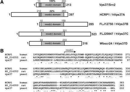
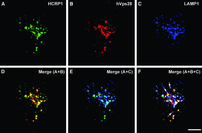
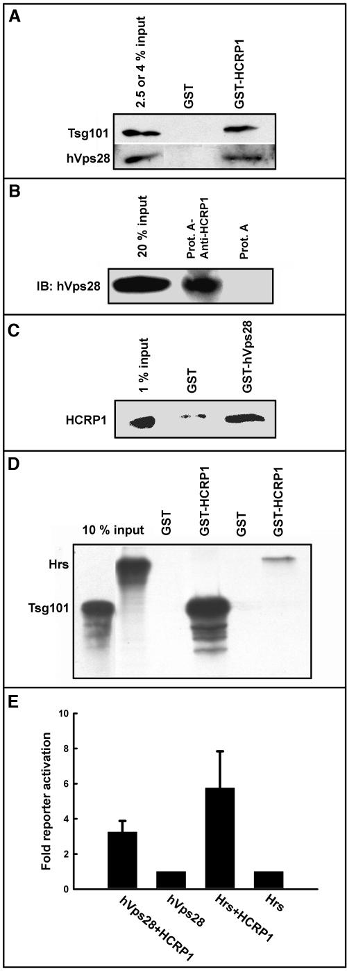
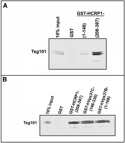
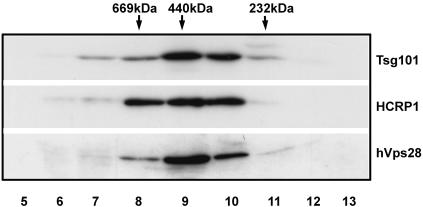
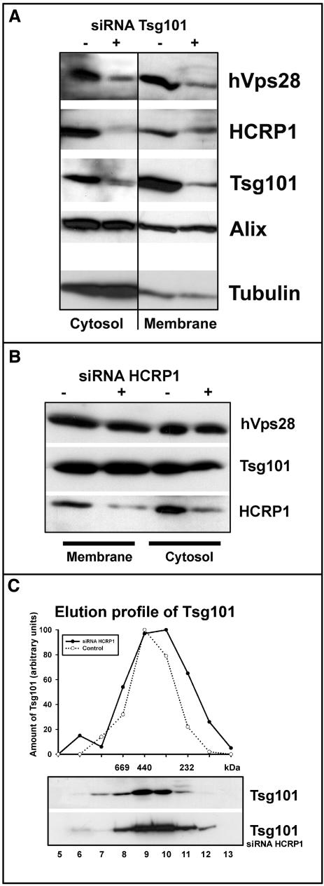
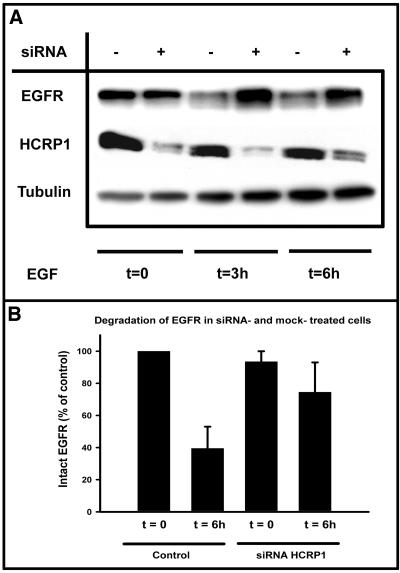
The biogenesis of multivesicular bodies and endosomal sorting of membrane cargo are driven forward by the endosomal sorting complexes required for transport, ESCRT-I, -II, and -III. ESCRT-I is characterized in yeast as a complex consisting of Vps23, Vps28, and Vps37. Whereas mammalian homologues of Vps23 and Vps28 (named Tsg101 and hVps28, respectively) have been identified and characterized, a mammalian counterpart of Vps37 has not yet been identified. Here, we show that a regulator of proliferation, hepatocellular carcinoma related protein 1 (HCRP1), interacts with Tsg101, hVps28, and their upstream regulator Hrs. The ability of HCRP1 (which we assign the alternative name hVps37A) to interact with Tsg101 is conferred by its mod(r) domain and is shared with hVps37B and hVps37C, two other mod(r) domain-containing proteins. HCRP1 cofractionates with Tsg101 and hVps28 by size exclusion chromatography and colocalizes with hVps28 on LAMP1-positive endosomes. Whereas depletion of Tsg101 by siRNA reduces cellular levels of both hVps28 and HCRP1, depletion of HCRP1 has no effect on Tsg101 or hVps28. Nevertheless, HCRP1 depletion strongly retards epidermal growth factor (EGF) receptor degradation. Together, these results indicate that HCRP1 is a subunit of mammalian ESCRT-I and that its function is essential for lysosomal sorting of EGF receptors.


Figures

Figure 1.
Figure 1.
HCRP1 is a possible mammalian counterpart of yeast Vps37. (A) Domain structure of Vps37 (NP_013220) and HCRP1 (AAK54349) is shown. The regions that are most conserved between these proteins are shaded, with the calculated sequence identity indicated. These conserved regions contain a modifier of rudimentary [mod(r)] domain (pfam: PF07200) (light gray), which is found in several eukaryotic proteins. These include three additional human protein products, FLJ20847 (hVps37C), FLJ12750 (hVps37B), and Wbscr24 (hVps37D), which are shown in the figure. The mod(r) domain of CAD38936 (the partial sequence of FLJ20847) includes the residues (26–175) (see Materials and Methods). (B) Alignment of the conserved region between Vps37 and HCRP1 is shown. The rat counterpart (XP_214353) of HCRP1 also is included. The starting point of the mod(r) domain is indicated.
Figure 2.
Figure 2.
HCRP1 and hVps28 colocalize on LAMP-1–containing endosomes. HeLa cells were cotransfected with hVps28 and GFP-HCRP1 for 24 h and permeabilized before fixation. They were labeled with anti-hVps28 (B) and anti-LAMP-1 (C). Colocalization between HCRP1 and hVps28 is shown in yellow (D), between LAMP-1 and HCRP1 in turquoise (E), and between all three molecules in white (F). Examples of profiles positive for all three molecules are indicated by arrows. Bar, 5 μm.
Figure 3.
Figure 3.
HCRP1 interacts with Tsg101, hVps28, and Hrs. (A) Interaction between HCRP1, Tsg101, and hVps28 in HeLa cells. GST alone or fused to HCRP1 was immobilized on glutathione-Sepharose beads and incubated with cell lysate from HeLa cells. The beads were washed and analyzed by SDS-PAGE and Western blotting with antibodies against Tsg101 and hVps28; 2.5 and 4% of the inputs of Tsg101 and hVps28, respectively, were loaded in lane 1. The amounts of Tsg101 and hVps28 pulled down with GST-HCRP1 correspond to 2–3 and 1–2% of the input amounts. (B) HCRP1 interacts with hVps28 in vivo. Protein A-Sepharose alone or bound to anti-HCRP1 was incubated with cell lysate from HeLa cells for 1 h at 4°C. The beads were washed and analyzed on SDS-PAGE and Western blotting with antibodies against hVps28. About 10% of hVps28 coimmunoprecipitated with HCRP1. (C) GST alone or fused to hVps28 was immobilized on glutathione-Sepharose beads and incubated with in vitro translated, 35S-labeled HCRP1 for 1 h at 4°C. The beads were then washed and analyzed by SDS-PAGE and fluorography. The amount bound corresponds to 1–2% of the input amount. (D) GST alone or fused to HCRP1 was immobilized on glutathione-Sepharose beads and incubated with in vitro translated, 35S-labeled Hrs or Tsg101 for 1 h at 4°C. The beads were washed and analyzed by SDS-PAGE and fluorography. (E) interactions of HCRP1 with hVps28 and Hrs in the yeast two-hybrid system. HCRP1 was used as prey and hVps28 and Hrs as baits. The values indicate β-galactosidase activities presented as fold reporter activation, and control bars (hVps28 and Hrs) represent background activation. The HCRP1 prey construct did not show any reporter activation in the absence of a bait construct (unpublished data).
Figure 4.
Figure 4.
The mod(r) domains of HCRP1 and hVps37B/C interact with Tsg101. (A) GST alone or fused with HCRP1(1–148) or HCRP1(208–397) was immobilized on glutathione-Sepharose beads and incubated with in vitro-translated 35S-labeled Tsg101 for 1 h at 4°C. The beads were then washed and analyzed by SDS-PAGE and fluorography. The input amount is indicated. (B) GST alone or fused with HCRP1 (208–397), hVps37C (146–330), or hVps37B (1–168) was immobilized on glutathione-Sepharose beads and incubated with in vitro-translated 35S-labeled Tsg101 as described in A. The doublet band representing Tsg101 is probably due to translational initiation at alternative positions in vitro (Bache et al., 2003).
Figure 5.
Figure 5.
Size exclusion chromatography of ESCRT-I. HeLa cells were lysed, followed by size exclusion chromatography. In total, 18 fractions were analyzed by SDS-PAGE and anti-Tsg101, HCRP1, and hVps28 blotting. Only fractions 5–13 are shown; no immunoreactivity was detected in the other fractions. The molecular mass standards and fraction numbers are indicated.
Figure 6.
Figure 6.
Depletion of Tsg101 by siRNA reduces cellular levels of hVps28 and HCRP1. (A) HeLa cells treated with control RNA (–)or with siRNA against Tsg101 (+) were fractionated into membrane and cytosolic fractions as described in Materials and Methods. Tsg101 left in the cytosol and on membranes after siRNA treatment as described in Materials and Methods was analyzed by SDS-PAGE, and the corresponding levels of hVps28, HCRP1, and Alix were shown by sequential blotting of the same membrane with anti-hVps28, anti-HCRP1, or anti-Alix. Equal amount of loaded protein is verified by blotting with anti-tubulin. (B) HeLa cells treated with control RNA (–) or with siRNA against HCRP1 (+) were fractionated into membrane and cytosolic fractions as described in Materials and Methods. The fractions were analyzed by Western blotting by using antibodies against HCRP1, Tsg101, and hVps28. (C) Lysates from HeLa cells treated with siRNA against HCRP1 (bottom gel) or not (top gel) were run on size exclusion chromatography. Eighteen fractions were analyzed by SDS-PAGE and Western blotting by using antibodies against Tsg101. Only fraction 5–13 are shown, because no Tsg101 was detected in any other fractions. The intensity of the bands was measured using ImageQuant 5.0 and plotted as percentage of the strongest band (top). Control cells, open symbols; HCRP1 siRNA-treated cells, closed symbols.
Figure 7.
Figure 7.
Depletion of HCRP1 retards EGF receptor down-regulation. (A) HeLa cells treated with control RNA (–) or with siRNA against HCRP1 (+) were stimulated for 0, 3, and 6 h with EGF. The cells were lysed and analyzed by SDS-PAGE and sequential blotting with antibodies against EGF receptor and HCRP1. The same membrane was then reblotted with anti-tubulin to verify equal loadings. (B) Relative intensities of the bands from control and siRNA-treated cells incubated with EGF for 0 or 6 h were quantified using Image-Quant 5.0 and are presented as the average of two experiments. Error bars denote SEM.

References

    1. Babst, M., Katzmann, D.J., Estepa-Sabal, E.J., Meerloo, T., and Emr, S.D. (2002a). Escrt-III: an endosome-associated heterooligomeric protein complex required for mvb sorting. Dev. Cell 3, 271–282. - PubMed
    1. Babst, M., Katzmann, D.J., Snyder, W.B., Wendland, B., and Emr, S.D. (2002b). Endosome-associated complex, ESCRT-II, recruits transport machinery for protein sorting at the multivesicular body. Dev. Cell 3, 283–289. - PubMed
    1. Babst, M., Odorizzi, G., Estepa, E.J., and Emr, S.D. (2000). Mammalian tumor susceptibility gene 101 (TSG101) and the yeast homologue, Vps23p, both function in late endosomal trafficking. Traffic 1, 248–258. - PubMed
    1. Bache, K.G., Brech, A., Mehlum, A., and Stenmark, H. (2003). Hrs regulates multivesicular body formation via ESCRT recruitment to endosomes. J. Cell Biol. 162, 435–442. - PMC - PubMed
    1. Begley, D., Murphy, A.M., Hiu, C., and Tsubota, S.I. (1995). Modifier of rudimentary p1, mod(r)p1, a trans-acting regulatory mutation of rudimentary. Mol. Gen. Genet. 248, 69–78. - PubMed

Publication types

MeSH terms