. 2004 Jul 13;101(28):10296-301.
doi: 10.1073/pnas.0403175101. Epub 2004 Jul 6.

The serine/threonine kinase cyclin G-associated kinase regulates epidermal growth factor receptor signaling

Affiliations

The serine/threonine kinase cyclin G-associated kinase regulates epidermal growth factor receptor signaling

Lei Zhang et al. Proc Natl Acad Sci U S A. .

Abstract

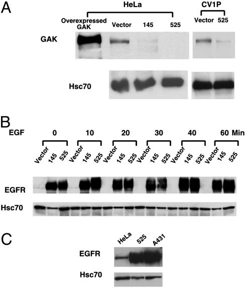
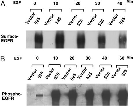
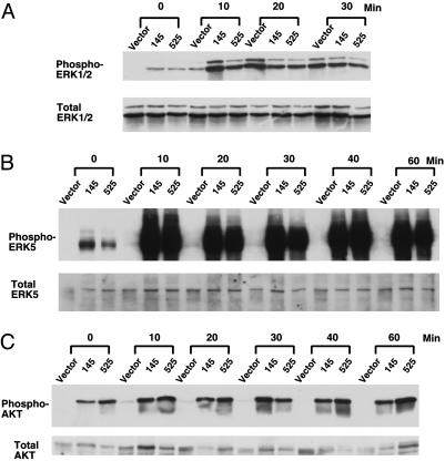
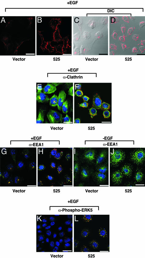
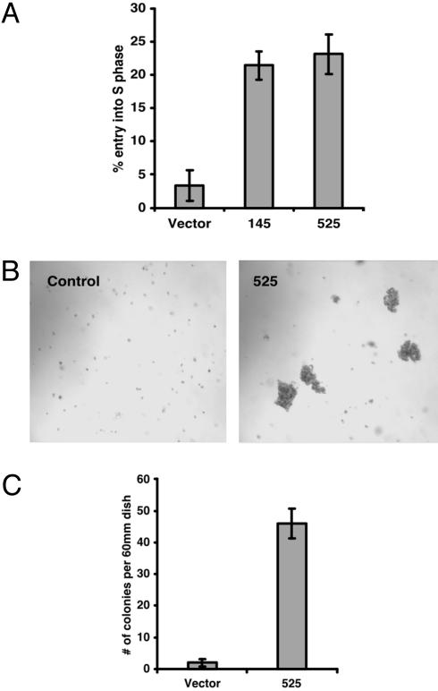
Cyclin G-associated kinase (GAK) is a serine/threonine kinase that features high homology outside its kinase domain with auxilin. Like auxilin, GAK has been shown to be a cofactor for uncoating clathrin vesicles in vitro. We investigated epidermal growth factor (EGF) receptor-mediated signaling in cells, in which GAK is down-regulated by small hairpin RNAs. Here, we report that down-regulation of GAK by small hairpin RNA has two pronounced effects on EGF receptor signaling: (i) the levels of receptor expression and tyrosine kinase activity go up by >50-fold; and (ii) the spectrum of downstream signaling is significantly changed. One very obvious result is a large increase in the levels of activated extracellular signal-regulated kinase 5 and Akt. These two effects of GAK down-regulation result from, at least in part, alterations in receptor trafficking, the most striking of which is the persistence of EGF receptor in altered cellular compartment along with activated extracellular signal-regulated kinase 5. The alterations resulting from GAK down-regulation can have distinctive biological consequences: In CV1P cells, down-regulation of GAK results in outgrowth of cells in soft agar, raising the possibility that loss of GAK function may promote tumorigenesis.


Figures

Fig. 1.
Fig. 1.
Down-regulation of GAK dramatically increases EGFR expression in HeLa cells. (A) GAK expression was stably down-regulated in HeLa and CV1P cells. Cells were infected with retroviral pSuper vectors encoding either one of two hairpin constructs capable of generating 19-nt duplex RNAi oligonucleotides corresponding to human GAK sequences, starting at the 145th base in the coding sequence (denoted 145) or the 525th base (denoted 525). Western blotting of total cell lysates was carried out by using a mAb against GAK. As a positive control, a lysate from cells transiently overexpressing GAK was included. Levels of Hsc70 were used as loading controls. (B) EGFR expression was dramatically enhanced in GAK knockdown cells. Cells were serum-starved overnight and stimulated with 50 ng/ml EGF for the indicated times. EGFR was detected by Western blotting using an Ab against it. (C) The level of EGFR expression in GAK knockdown cells was ≈75% of that of A431 cells. Cells were serum-starved overnight and EGFR expression was detected as described in B. Levels of Hsc70 were used as loading controls.
Fig. 2.
Fig. 2.
In GAK knockdown cells, EGFR remains surface-associated for extended periods after EGF stimulation and displays enhanced kinase activity. (A) The level of surface-associated EGFR in GAK knockdown cell remains high for a prolonged time. Cells were serum-starved overnight and incubated with 50 ng/ml EGF for indicated times. Cells were surface-labeled with biotin at the indicated times, then the biotinylated receptors were isolated by means of streptavidin affinity and visualized by Western blotting with an EGFR Ab. (B) EGFR in GAK knockdown cells displays high tyrosine kinase activity in response to EGF stimulation. Cells were serum-starved overnight and incubated with 50 ng/ml EGF for indicated times. Total EGFR in cells was immunoprecipitated with EGFR Ab and blotted with an Ab against phosphorylated tyrosine (4G10).
Fig. 3.
Fig. 3.
Down-regulation of GAK differentially enhances EGFR-dependent signaling. Cells were serum-starved overnight and stimulated with 50 ng/ml EGF for indicated times. Western blot analysis was carried out by probing with an Ab against phosphorylated ERK1/2 or total ERK1/2(A); and an Ab against phosphorylated ERK5 or total ERK5 (B); or an Ab against phosphorylated Akt or total Akt (C).
Fig. 4.
Fig. 4.
Down-regulation of GAK alters intracellular trafficking of EGFR. Cells grown on chamber slides were serum-starved overnight. Cells were then incubated with 0.5 μg/ml rhodamine-conjugated EGF on ice for 1 h. (AD) Cells were either fixed immediately (A and B) or incubated at 37°C in a 10% CO2 humidified incubator for 40 min (C and D), then fixed and counterstained with DAPI (blue) to localize the nucleus of the cell being studied. (E–L) Alternatively, cells were incubated with (E–H, K, and L) or without (I and J) rhodamineconjugated EGF (red signal) and fixed as described above, then incubated with primary Ab as indicated below. In each case, the Ab signal is green. Overlap of the EGF signal in red and Ab signal in green is yellow. Cells were visualized with a Zeiss LSM 510 META/NLO confocal microscope at ×63 magnification and images were captured by Zeiss confocal microscope software Version 3.2. (A and B) Enhanced levels of EGFR visualized on the surface of GAK knockdown cells. Fluorescently labeled EGF is used to visualize EGFR on the plasma membrane without internalization. (C and D) EGFR is mislocalized after EGF stimulation. Fluorescently labeled EGF is used to visualize EGFR at 40 min after stimulation. Images captured by using differential interference contrast (DIC) and fluorescence microscopy combined. (E and F) EGFR partially colocalizes with clathrin. Costaining with an Ab against clathrin in addition to fluorescently labeled EGF is used to determine the localization of clathrin in the compartment containing the EGFR. (G and H) EGFR colocalizes with EEA1 in GAK knockdown cells. Costaining with an Ab against EEA1, an early endosome marker, in addition to fluorescently labeled EGF is used to determine the degree of colocalization between EEA1 and EGFR. (I and J) EEA1 is mislocalized even in the absence of receptor stimulation. An Ab against EEA1 is used to show the status of the early endosome in the absence of EGF stimulation. (K and L) Activated ERK5 colocalizes with EGFR. Costaining with an Ab against phosphorylated ERK5 in addition to fluorescently labeled EGF is used to show the relationship between EGFR and activated ERK5. (Scale bar, 20 μM.)
Fig. 5.
Fig. 5.
Down-regulation of GAK promotes cell proliferation and cell transformation. (A) Down-regulation of GAK promotes cell proliferation under conditions of serum starvation. Cells grown on chamber slides were serumstarved overnight and labeled with BrdUrd for 15 h. Cells were fixed and incubated with an Ab against BrdUrd. BrdUrd-positive cells were counted among DAPI-stained cells in at least 10 randomly selected fields on each slide (n = 3). (B) Down-regulation of GAK transforms CV1P cells. CV1P cells (vector control and 525) were seeded in soft agar. Phase-contrast image of the colonies from a representative experiment is shown (magnification: ×7). (C) Colony numbers (colony with >50 cells) from B were counted at 21 days after cells were seeded (n = 3).

References

    1. Kanaoka, Y., Kimura, S. H., Okazaki, I., Ikeda, M. & Nojima, H. (1997) FEBS Lett. 402, 73–80. - PubMed
    1. Kimura, S. H., Tsuruga, H., Yabuta, N., Endo, Y. & Nojima, H. (1997) Genomics 44, 179–187. - PubMed
    1. Greener, T., Zhao, X., Nojima, H., Eisenberg, E. & Greene, L. E. (2000) J. Biol. Chem. 275, 1365–1370. - PubMed
    1. Umeda, A., Meyerholz, A. & Ungewickell, E. (2000) Eur. J. Cell Biol. 79, 336–342. - PubMed
    1. Korolchuk, V. I. & Banting, G. (2002) Traffic 3, 428–439. - PubMed

Publication types

MeSH terms