. 2004 Jul 13;101(28):10278-83.
doi: 10.1073/pnas.0401911101. Epub 2004 Jul 6.

Template-assisted filament growth by parallel stacking of tau

Affiliations

Template-assisted filament growth by parallel stacking of tau

Martin Margittai et al. Proc Natl Acad Sci U S A. .

Abstract

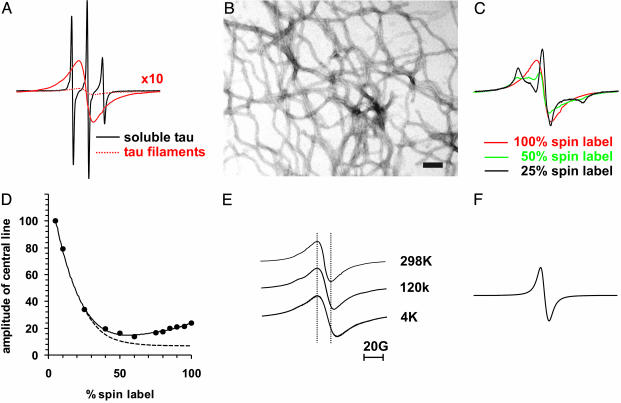
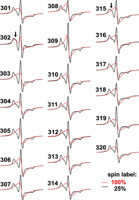
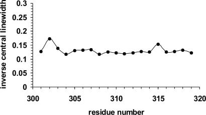
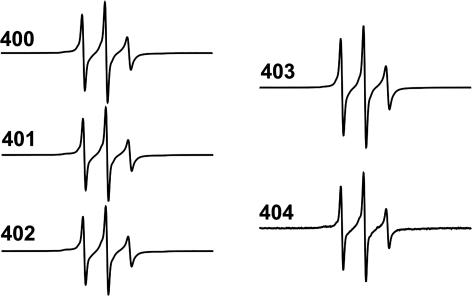
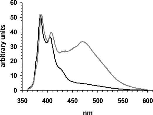
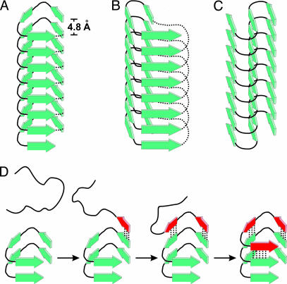
Tau filaments are found in >20 neurodegenerative diseases. Yet, because of their enormous molecular weights and poor tendency to form highly ordered 3D crystal lattices, they have evaded high-resolution structure determination. Here, we studied 25 derivatized tau mutants by using electron paramagnetic resonance and fluorescence spectroscopy to report structural details of tau filaments. Based on strong spin exchange and pyrene excimer formation of core residues, we find that individual tau proteins form single molecule layers along the fiber axis that perfectly stack on top of each other by in-register, parallel alignment of beta-strands. We suggest a model of filament growth wherein the existing filament serves as a template for the incoming, unfolded tau molecule, resulting in a new structured layer with maximized hydrogen-bonded contact surface and side-chain stacking.


Figures

Fig. 1.
Fig. 1.
Bar diagram of the microtubule-associated protein tau. The large isoform of tau (441 aa) contains two inserts in the near N-terminal half (I1 and I2, 29 aa) and four microtubule-binding regions (R1–R4, 31–32 aa) in the C-terminal half. Sites that were investigated in this study are depicted by single-letter amino acid code.
Fig. 2.
Fig. 2.
Spin exchange in a core residue of tau. Tau spectra and EM micrograph were taken from a cysteine mutant labeled at position 308. (A) Overlay of spectra from soluble tau (black), and filamentous tau (dotted red), for better comparison the amplitude of the spectrum of filamentous tau is multiplied by 10 (red line). (B) EM micrograph (×50,000) of uranyl acetate stained tau filaments. (Bar = 100 nm.) (C) Spectra of tau filaments containing 100% spin label (red as in A), 50% spin label (green), and 25% spin label (black); see also Materials and Methods. (D) Amplitudes of central lines from various dilutions are plotted vs. the percentage of spin label. Dotted line schematically illustrates decrease in amplitude as observed for dipolar coupling (see, e.g., refs. and 49). (E) Spectra from filaments taken at 298, 120, and 4 K. Spectra are normalized to the same amplitude. (F) Spectrum of crystals from MTSL spin label.
Fig. 3.
Fig. 3.
EPR spectra of spin-labeled positions 301–320 in tau filaments. Spectra of tau filaments labeled with 100% spin label (red) or 25% spin label (black). Arrows point to more mobile components in the dilution spectra of positions 302 and 315.
Fig. 4.
Fig. 4.
Inverse linewidth plot. The inverse central linewidths from dilution spectra of tau filaments are plotted against residue number.
Fig. 5.
Fig. 5.
EPR spectra of spin-labeled positions 400–404 in the tau filament.
Fig. 6.
Fig. 6.
Excimer formation. Emission spectra (360–600 nm) from tau filaments derivatized at positions 308 (gray) and 403 (black) normalized to the 385-nm peak. Samples were excited at 344 nm.
Fig. 7.
Fig. 7.
Substructure of tau filament and layer extension. β-Strands in tau filaments have parallel, in-register alignment and are perfectly stacked along the fiber axis. Individual layers of tau molecules are hydrogen bonded and separated by 4.8 Å. (AC) Models of tau filaments with different possible lateral arrangements are depicted: left-handed β-helix (A), right-handed β-helix (B), and intersheet-hairpin (C). Dotted lines represent intramolecular connections in β-helices of soluble proteins as observed in crystal structures (39). In tau filaments this continuity would be disrupted and neighboring repeat regions would project from both ends, possibly forming individual domains. (D) Filament growth is, for convenience, demonstrated on the left-handed “helix” model. The incoming unstructured tau molecule forms β-strands (red) and hydrogen bonds (dotted lines) to a filament template adding one new layer through a zipper-like mechanism.

References

    1. Lee, V. M., Goedert, M. & Trojanowski, J. Q. (2001) Annu. Rev. Neurosci. 24, 1121–1159. - PubMed
    1. Goedert, M., Spillantini, M. G., Cairns, N. J. & Crowther, R. A. (1992) Neuron 8, 159–168. - PubMed
    1. Metuzals, J., Montpetit, V. & Clapin, D. F. (1981) Cell Tissue Res. 214, 455–482. - PubMed
    1. Yagishita, S., Itoh, Y., Nan, W. & Amano, N. (1981) Acta Neuropathol. 54, 239–246. - PubMed
    1. Wischik, C. M., Novak, M., Edwards, P. C., Klug, A., Tichelaar, W. & Crowther, R. A. (1988) Proc. Natl. Acad. Sci. USA 85, 4884–4888. - PMC - PubMed

Publication types

LinkOut - more resources