. 2004 Jul 13;101(28):10254-9.
doi: 10.1073/pnas.0400014101. Epub 2004 Jul 6.

Insight into the PrPC-->PrPSc conversion from the structures of antibody-bound ovine prion scrapie-susceptibility variants

Affiliations

Insight into the PrPC-->PrPSc conversion from the structures of antibody-bound ovine prion scrapie-susceptibility variants

Frédéric Eghiaian et al. Proc Natl Acad Sci U S A. .

Abstract

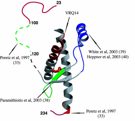
Prion diseases are associated with the conversion of the alpha-helix rich prion protein (PrPC) into a beta-structure-rich insoluble conformer (PrPSc) that is thought to be infectious. The mechanism for the PrPC-->PrPSc conversion and its relationship with the pathological effects of prion diseases are poorly understood, partly because of our limited knowledge of the structure of PrPSc. In particular, the way in which mutations in the PRNP gene yield variants that confer different susceptibilities to disease needs to be clarified. We report here the 2.5-A-resolution crystal structures of three scrapie-susceptibility ovine PrP variants complexed with an antibody that binds to PrPC and to PrPSc; they identify two important features of the PrPC-->PrPSc conversion. First, the epitope of the antibody mainly consists of the last two turns of ovine PrP second alpha-helix. We show that this is a structural invariant in the PrPC-->PrPSc conversion; taken together with biochemical data, this leads to a model of the conformational change in which the two PrPC C-terminal alpha-helices are conserved in PrPSc, whereas secondary structure changes are located in the N-terminal alpha-helix. Second, comparison of the structures of scrapie-sensitivity variants defines local changes in distant parts of the protein that account for the observed differences of PrPC stability, resistant variants being destabilized compared with sensitive ones. Additive contributions of these sensitivity-modulating mutations to resistance suggest a possible causal relationship between scrapie resistance and lowered stability of the PrP protein.


Figures

Fig. 1.
Fig. 1.
Structure of the OvPrP C-terminal domain. (A) The OvPrP fold and the scrapie-sensitivity-associated mutations. Side chains of scrapie-sensitivity-related residues are represented as green ball-and-stick models in the OvPrP structure. (B Left) Structure of the huPrP crystallographic dimer (27) (one monomer is shown in salmon red, and the other is shown in blue). (B Right) Superimposition of the x-ray structures of OvPrP (this work, red) and of a huPrP domain (blue) constituted by the H3 helix of one monomer and of the rest of the sequence of the other monomer.
Fig. 2.
Fig. 2.
Structural consequences of scrapie-sensitivity-related mutations. The blue grid corresponds to the 1.0-σ level contour of the 2Fo – Fc omit map. Maps are calculated by using data in the 15.0- to 2.5-Å-resolution range for the ARQ and VRQ variants and the 15.0- to 2.8-Å-resolution range for the ARR variant. Hydrogen bonds are displayed as dashed lines, and distances of the atoms involved are reported. (Upper) A136V. The FoVRQ – FoARQ difference map, represented as red and green grids (contour levels: –5.0 σ and 5.0 σ, respectively) is superimposed on the ARQ 2Fo – Fc omit map. (Lower) Q171R.
Fig. 3.
Fig. 3.
The epitope of the antibody is conserved in OvPrPC and OvPrPSc. (A) OvPrP–Fab structure (ARQ variant). Fab heavy and light chains are shown in green and blue, respectively. (Left) Overview of the complex. The part of the prion protein that, according to our model, is structurally conserved in the PrPC → PrPSc conversion is shown in red; the part that undergoes a secondary structure change is shown in orange. (Right) Close-up view of the boxed region of the complex. For clarity, only atoms that establish an intermolecular hydrogen bond are represented in ball-and-stick format. Hydrogen bonds are represented as dashed lines, and distances of the atoms involved (Å) are reported. (B) ELISA characterization of the binding properties of the antibody. PrPC and PrPC + PrPSc samples are recovered from brains of uninfected and infected sheep, respectively. PrPSc is produced by proteinase K (PK) treatment of PrPC + PrPSc. The proteinase K treatment of PrPC samples is used as a control for total digestion of PrPC. Prnp0/0 designates an extract from the brain of a Prnp0/0 mouse. (Upper) ELISA titration of OvPrP samples. (Lower) Competition ELISA of the Fab and the antibody. The lower signal for PrPSc in both the titration and the competition tests reflects the lower abundance of PrPSc compared with PrPC in infected sheep brains (–43). A.U., arbitrary units.
Fig. 4.
Fig. 4.
Antigenic mapping of the structural changes in the PrPC → PrPSc conversion. The epitopes are mapped on the OvPrP structure to which three residues have been added at the C terminus; their conformation is that determined by NMR. The epitopes of PrPSc-specific antibodies are shown in green, and those present in both PrPC and PrPSc are shown in red. The conserved disulfide is shown in red also. Antibody binding to the region in blue blocks PrPSc formation in vivo. Each displayed epitope is linked to its bibliographic input.

References

    1. Prusiner, S. B. (1998) Proc. Natl. Acad. Sci. USA 95, 13363–13383. - PMC - PubMed
    1. Bueler, H., Aguzzi, A., Sailer, A., Greiner, R. A., Autenried, P., Aguet, M. & Weissmann, C. (1993) Cell 73, 1339–1347. - PubMed
    1. Lasmezas, C. I., Deslys, J. P., Robain, O., Jaegly, A., Beringue, V., Peyrin, J. M., Fournier, J. G., Hauw, J. J., Rossier, J. & Dormont, D. (1997) Science 275, 402–405. - PubMed
    1. Goldmann, W., Hunter, N., Foster, J. D., Salbaum, J. M., Beyreuther, K. & Hope, J. (1990) Proc. Natl. Acad. Sci. USA 87, 2476–2480. - PMC - PubMed
    1. Goldmann, W., Martin, T., Foster, J., Hughes, S., Smith, G., Hughes, K., Dawson, M. & Hunter, N. (1996) J. Gen. Virol. 77, 2885–2891. - PubMed

Publication types