. 2004 Jul 13;101(28):10482-7.
doi: 10.1073/pnas.0403930101. Epub 2004 Jul 6.

Lipid- and mechanosensitivities of sodium/hydrogen exchangers analyzed by electrical methods

Affiliations

Lipid- and mechanosensitivities of sodium/hydrogen exchangers analyzed by electrical methods

Daniel Fuster et al. Proc Natl Acad Sci U S A. .

Abstract

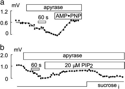
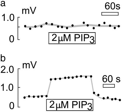
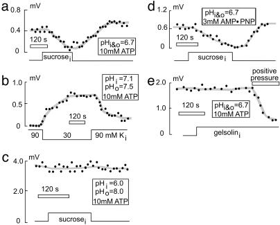
Sodium/hydrogen exchangers (NHEs) are ubiquitous ion transporters that serve multiple cell functions. We have studied two mammalian isoforms, NHE1 (ubiquitous) and NHE3 (epithelial-specific), by measuring extracellular proton (H+) gradients during whole-cell patch clamp with perfusion of the cell interior. Maximal Na(+)-dependent H+ fluxes (JH+) are equivalent to currents >20 pA for NHE1 in Chinese hamster ovary fibroblasts, >200 pA for NHE1 in guinea pig ventricular myocytes, and 5-10 pA for NHE3 in opossum kidney cells. The fluxes are blocked by an NHE inhibitor, ethylisopropylamiloride, and are absent in NHE-deficient AP-1 cells. NHE1 activity is stable with perfusion of nonhydrolyzable ATP [adenosine 5'-(beta,gamma-imido)triphosphate], is abolished by ATP depletion (2 deoxy-D-glucose with oligomycin or perfusion of apyrase), can be restored with phosphatidylinositol 4,5-bisphosphate, and is unaffected by actin cytoskeleton disruption (latrunculin or pipette perfusion of gelsolin). NHE3 (but not NHE1) is reversibly activated by phosphatidylinositol 3,4,5-trisphosphate. Both NHE1 and NHE3 activities are disrupted in giant patches during gigaohm seal formation. NHE1 (but not NHE3) is reversibly activated by cell shrinkage, even at neutral cytoplasmic pH without ATP, and inhibited by cell swelling. NHE1 in Chinese hamster ovary fibroblasts (but not NHE3 in opossum kidney cells) is inhibited by agents that thin the membrane (L-alpha-lysophosphatidylcholine and octyl-beta-D-glucopyranoside) and activated by cholesterol enrichment, which thickens membranes. Expressed in AP-1 cells, however, NHE1 is insensitive to these agents but remains sensitive to volume changes. Thus, changes of hydrophobic mismatch can modulate NHE1 but do not underlie its volume sensitivity.


Figures

Fig. 2.
Fig. 2.
Effect of ATP depletion on NHE1 activity in CHO cells. (a and b) Pipette perfusion with apyrase (5 units/ml) leads to a rapid decline in NHE1 activity in CHO cells. Perfusion of 10 mM AMP-PNP in the presence of apyrase (5 units/ml) and 2 mM EDTA leads to a complete recovery of NHE1 activity (a), whereas perfusion with PIP2 only partially recovers NHE1 activity (b). Even in the absence of ATP, NHE1 activity remains volume-sensitive (b).
Fig. 3.
Fig. 3.
Effect of PIP3 on NHE activity. Shown is pipette perfusion of PIP3 into CHO (a) and OK (b) cells. NHE3 but not NHE1 activity is stimulated rapidly and reversibly by 2 μM cytoplasmic PIP3.
Fig. 4.
Fig. 4.
Effect of cell-volume changes on NHE1 activity in CHO cells. Pipette perfusion with a hypertonic solution containing sucrose leads to cell swelling and reduction of NHE1 activity. (a) This is reversed by perfusion of hypotonic solution without sucrose, which restores cell volume. (b) Vice versa, perfusion of hypotonic solution leads to cell swelling and increase NHE1 activity, even under conditions of a normal pHi of 7.1. (c) At maximal activation of NHE1 activity (pHi 6 and pHo 8), volume sensitivity of NHE1 is lost. (d) In the absence of MgATP in the pipette solution and replacement by AMP-PNP, volume sensitivity of NHE1 is preserved. (e) Pipette perfusion with gelsolin to depolymerize F-actin does not affect NHE1 activity or NHE1 volume sensitivity.
Fig. 5.
Fig. 5.
Effect of bilayer modulation by hydrophobic compounds on NHE1 activity. Pipette perfusion (a) or bath application (c) of 1 mM OG strongly inhibits NHE1 activity in CHO cells but does not affect NHE3 activity in OK cells (c). (c) Similar effects occur with pipette perfusion of 10 μM l-α-lysophosphatidylcholine (LPC). Pipette perfusion of 100 μM cholesterol complexed with MCD strongly stimulates NHE1 activity in CHO cells (b) but does not affect NHE3 activity in OK cells (c). (c) Cholesterol depletion with pipette perfusion of 1 mM MCD does not affect either NHE1 or NHE3 activity. (d) NHE1 transfected into AP-1 cells is not affected by pipette perfusion of 1 mM OG or 100 μM cholesterol.
Fig. 1.
Fig. 1.
NHE activity assayed by pH microelectrodes: typical extracellular pH gradients detected by pH microelectrodes during whole-cell patch clamp recording. (a) Schematic diagram of the experimental procedure. Relative motion of the cell to the pH microelectrode spans ≈50 μM (arrow). (bk) Experimental records in which a cell was moved repetitively close to and away from (black bars) the pH microelectrode. Extracellular Na+ is 140 mM and pH is 6.7 on both sides unless indicated otherwise. (b) H+ gradient next to a CHO cell; upward deflection denotes higher pH. (c) H+ gradient next to a CHO cell with pHo 8.0 and pHi 6.0. Complete lack of H+ gradient next to a CHO cell is shown in d (in the presence of 10 μM ethylisopropylamiloride and 30 mM Na+) and e (with extracellular Na+ replaced by K+). (f) Complete lack of a pH gradient next to an NHE-deficient AP-1 cell. Shown are pH gradients next to a guinea pig ventricular myocyte (g), an OK cell (h), and an AP-1 cell transfected with either wild-type (i) or different C-terminal truncated forms [j (NHE3 Δ1–462) and k (NHE3 Δ1–552)] of opossum NHE3.

References

    1. Orlowski, J. & Grinstein, S. (2003) Pflugers Arch. 447, 549–565. - PubMed
    1. Denker, S. P. & Barber, D. L. (2002) J. Cell Biol. 159, 1087–1096. - PMC - PubMed
    1. Putney, L. K. & Barber, D. L. (2003) J. Biol. Chem. 278, 44645–44649. - PubMed
    1. Putney, L. K., Denker, S. P. & Barber, D. L. (2002) Annu. Rev. Pharmacol. Toxicol. 42, 527–552. - PubMed
    1. Orlowski, J. & Grinstein, S. (1997) J. Biol. Chem. 272, 22373–22376. - PubMed

Publication types

Substances