. 2004 Jul 21;23(14):2821-9.
doi: 10.1038/sj.emboj.7600297. Epub 2004 Jul 8.

GRK2 is an endogenous protein inhibitor of the insulin signaling pathway for glucose transport stimulation

Affiliations

GRK2 is an endogenous protein inhibitor of the insulin signaling pathway for glucose transport stimulation

Isao Usui et al. EMBO J. .

Abstract

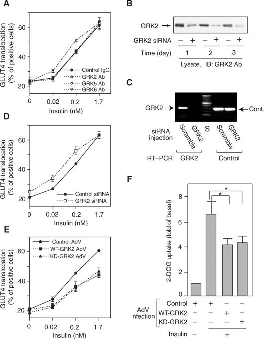
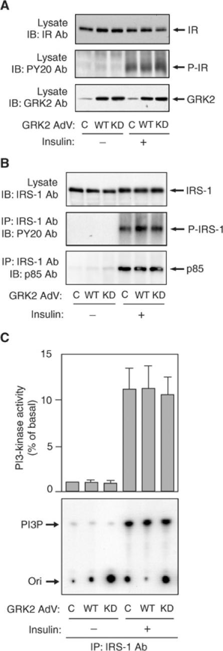
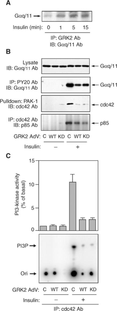
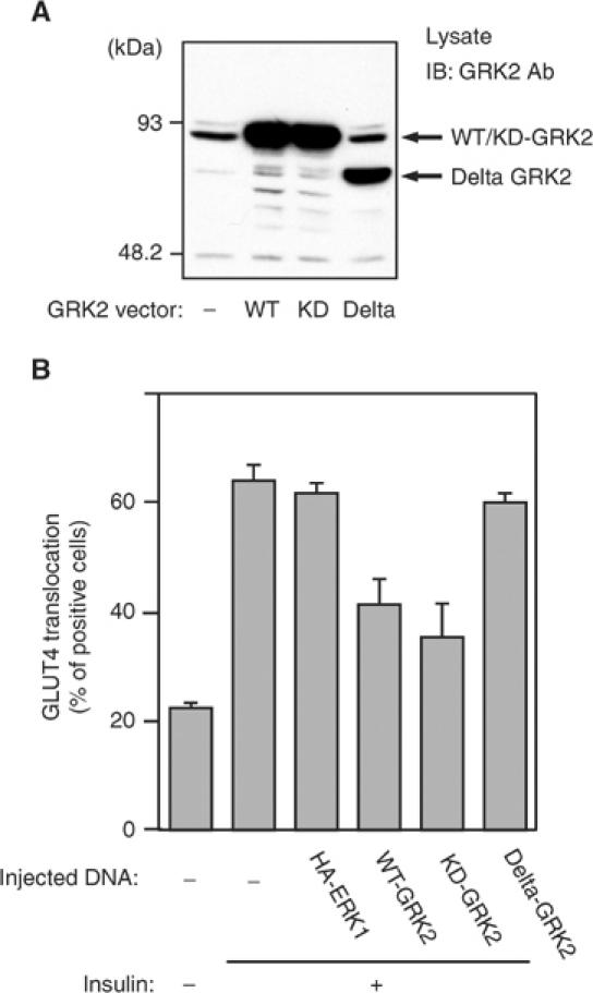
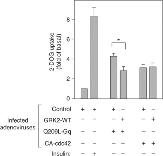
G protein-coupled receptor kinases (GRKs) represent a class of proteins that classically phosphorylate agonist-activated G protein-coupled receptors, leading to uncoupling of the receptor from further G protein activation. Recently, we have reported that the heterotrimeric G protein alpha-subunit, Galphaq/11, can mediate insulin-stimulated glucose transport. GRK2 contains a regulator of G protein signaling (RGS) domain with specificity for Galphaq/11. Therefore, we postulated that GRK2 could be an inhibitor of the insulin signaling cascade leading to glucose transport in 3T3-L1 adipocytes. In this study, we demonstrate that microinjection of anti-GRK2 antibody or siRNA against GRK2 increased insulin-stimulated insulin-responsive glucose transporter 4 (GLUT4) translocation, while adenovirus-mediated overexpression of wild-type or kinase-deficient GRK2 inhibited insulin-stimulated GLUT4 translocation as well as 2-deoxyglucose uptake. Importantly, a mutant GRK2 lacking the RGS domain was without effect. Taken together, these results indicate that through its RGS domain endogenous GRK2 functions as a negative regulator of insulin-stimulated glucose transport by interfering with Galphaq/11 signaling to GLUT4 translocation. Furthermore, inhibitors of GRK2 can lead to enhanced insulin sensitivity.


Figures

Figure 1
Figure 1
Effects of GRK2 on insulin-stimulated GLUT4 translocation and 2-DOG uptake in 3T3-L1 adipocytes. (A, D, E) For microinjection assay, 3T3-L1 adipocytes on coverslips were serum starved for 4 h, and anti-GRK2 antibody, anti-GRK5 antibody, anti-GRK6 antibody, sheep IgG, GRK2 siRNA, or control siRNA was microinjected. For GRK2 overexpression assay, 3T3-L1 adipocytes on coverslips were infected with adenovirus expressing WT-GRK2, KD-GRK2, or control LacZ. At 48 h after infection, these cells were serum starved for 4 h. Cells were stimulated with 0.02, 0.2, or 1.7 nM insulin for 20 min. GLUT4 was stained as described in Materials and methods. The percentage of cells positive for GLUT4 translocation was calculated by counting at least 100 cells at each point. The data are the mean±s.e. from three independent experiments. (B) SiRNA of GRK2 (+) or control siRNA (−) was transfected in 3T3-L1 preadipocytes using Oligofectamine as described in Materials and methods. At the indicated days after transfection, total cell lysates were prepared and analyzed by Western blotting using anti-GRK2 antibody as described in Materials and methods. Representative blots are shown from two independent experiments. (C) The efficiency of siRNA under the microinjection into 3T3-L1 adipocytes was confirmed by the mRNA quantification as described in Materials and methods. All of 3T3-L1 adipocytes on coverslips (approximately 200 cells spotted on each coverslip) were microinjected with GRK2 or scrambled control (scramble) siRNA. At 48 h after microinjection, total RNA was purified and used for RT–PCR reaction with the GRK2 or PP2A (Cont.) primer set. A representative image from two independent experiments is shown. S: size marker. (F) 3T3-L1 adipocytes were infected with adenovirus expressing WT-GRK2, KD-GRK2, or control LacZ (Control). At 48 h after infection, these cells were serum starved for 3 h, stimulated with17 nM insulin for 30 min, and [3H]2-DOG uptake was measured as described in Materials and methods. The data are the mean±s.e. from three independent experiments. Statistically significant differences versus control are indicated (*P<0.05).
Figure 2
Figure 2
Effect of WT- or KD-GRK2 overexpression on insulin receptor and IRS-1-PI3-kinase pathway in 3T3-L1 adipocytes. 3T3-L1 adipocytes were infected with adenovirus expressing WT-GRK2 (WT), KD-GRK2 (KD), or control LacZ (C). (A) At 48 h after infection, these cells were serum starved for 16 h, stimulated with 17 nM insulin for 5 min and lysed. Total cell lysates were analyzed by Western blotting using anti-insulin receptor (IR), anti-phosphotyrosine (PY20), or anti-GRK2 antibody as described in Materials and methods. Representative blots are shown from three independent experiments. (B) At 48 h after infection, these cells were serum starved for 16 h, stimulated with 17 nM insulin for 5 min (for IRS-1 and PY20 blots) or 10 min (for p85 blot) and lysed. Samples were immunoprecipitated with anti-IRS-1 antibody. Immunoprecipitates or total cell lysates were analyzed by Western blotting using anti-IRS-1, anti-phosphotyrosine (PY20), or anti-p85 antibody as described in Materials and methods. Representative blots are shown from three independent experiments. (C) At 48 h after infection, these cells were serum starved for 16 h, stimulated with 17 nM insulin for 10 min and lysed. Samples were immunoprecipitated with anti-IRS-1 antibody. PI3-kinase activity was measured as described in Materials and methods. A representative film is shown and the graph represents the mean±s.e. from three independent experiments.
Figure 3
Figure 3
Effect of WT- or KD-GRK2 overexpression on Gαq/11-cdc42-PI3-kinase pathway in 3T3-L1 adipocytes. (A) 3T3-L1 adipocytes were serum starved for 16 h, stimulated with 17 nM insulin for the indicated time periods and lysed. Samples were immunoprecipitated with anti-GRK2 antibody. Immunoprecipitates were analyzed by Western blotting using anti-Gαq/11 antibody as described in Materials and methods. A representative blot is shown from three independent experiments. (B) 3T3-L1 adipocytes were infected with adenovirus expressing WT-GRK2 (WT), KD-GRK2 (KD), or control LacZ (C). At 48 h after infection, these cells were serum starved for 16 h, stimulated with 17 nM insulin for 1 min (for Gαq/11 blot) or 10 min (for p85 blot) and lysed. Samples were immunoprecipitated with anti-phosphotyrosine (PY20) or cdc42 antibody. Immunoprecipitates and total cell lysates were analyzed by Western blotting using anti-Gαq/11 or anti-p85 antibody as described in Materials and methods. Cdc42 activity was measured as described in Materials and methods. The cells were stimulated with insulin for 1 min. Samples were pulled down with GST-PAK-1 and were analyzed by Western blotting using anti-cdc42 antibody. Representative blots are shown from three independent experiments. (C) 3T3-L1 adipocytes were infected with adenovirus expressing WT-GRK2 (WT), KD-GRK2 (KD), or control LacZ (C). At 48 h after infection, these cells were serum starved for 16 h, stimulated with 17 nM insulin for 10 min and lysed. Samples were immunoprecipitated with anti-cdc42 antibody. PI3-kinase activity was measured as described in Materials and methods. A representative film is shown and the graph represents the mean±s.e. from three independent experiments.
Figure 4
Figure 4
Effects of the RGS domain of GRK2 on insulin-stimulated GLUT4 translocation in 3T3-L1 adipocytes. (A) Plasmid expression vectors of WT-, KD-, or deletion mutant (Delta) GRK2 were transfected in HIRc-B cells using SuperFECT as described in Materials and methods. At 48 h after transfection, these cells were lysed and total cell lysates were analyzed by Western blotting using anti-GRK2 antibody as described in Materials and methods. These experiments were performed three times, and a representative result is shown. (B) Plasmid expression vectors of WT-GRK2, KD-GRK2, delta-GRK2, or control ERK1 (HA-ERK1) were microinjected into the nuclei of 3T3-L1 adipocytes on coverslips. At 24 h after microinjection, 3T3-L1 adipocytes were serum starved for 4 h and were stimulated with 1.7 nM insulin for 20 min. GLUT4 was stained as described in Materials and methods. The cells expressing exogenous GRKs or ERK1 were detected by staining with anti-6X-His antibody or anti-HA antibody, respectively. The percentage of cells positive for GLUT4 translocation was calculated by counting at least 100 cells at each point. The data are the mean±s.e. from three independent experiments.
Figure 5
Figure 5
Effects of GRK2 on Q209L-Gq-induced 2-DOG uptake in 3T3-L1 adipocytes. 3T3-L1 adipocytes were co-infected with adenovirus expressing WT-GRK2 (GRK2-WT), constitutively active Gq (Q209L-Gq), constitutively active cdc42 (CA-cdc42), and/or control LacZ (Control). Total number of MOI was adjusted to 80 in each condition. At 48 h after infection, these cells were serum starved for 3 h, stimulated with17 nM insulin for 30 min, and [3H]2-DOG uptake was measured as described in Materials and methods. The data are the mean±s.e. from six independent experiments. Statistically significant differences versus control are indicated (*P<0.05).

Similar articles

Cited by

References

    1. Baltensperger K, Karoor V, Paul H, Ruoho A, Czech MP, Malbon CC (1996) The beta-adrenergic receptor is a substrate for the insulin receptor tyrosine kinase. J Biol Chem 271: 1061–1064 - PubMed
    1. Carman CV, Parent JL, Day PW, Pronin AN, Sternweis PM, Wedegaertner PB, Gilman AG, Benovic JL, Kozasa T (1999) Selective regulation of Galpha(q/11) by an RGS domain in the G protein-coupled receptor kinase, GRK2. J Biol Chem 274: 34483–34492 - PubMed
    1. Chen JF, Guo JH, Moxham CM, Wang HY, Malbon CC (1997) Conditional, tissue-specific expression of Q205L G alpha i2 in vivo mimics insulin action. J Mol Med 75: 283–289 - PubMed
    1. Dalle S, Imamura T, Rose DW, Worrall DS, Ugi S, Hupfeld CJ, Olefsky JM (2002) Insulin induces heterologous desensitization of G-protein-coupled receptor and insulin-like growth factor I signaling by downregulating beta-arrestin-1. Mol Cell Biol 22: 6272–6285 - PMC - PubMed
    1. Dalle S, Ricketts W, Imamura T, Vollenweider P, Olefsky JM (2001) Insulin and insulin-like growth factor I receptors utilize different G protein signaling components. J Biol Chem 276: 15688–15695 - PubMed

Publication types

MeSH terms