. 2004 Jul;15(7):681-90.
doi: 10.1089/1043034041361271.

Novel bidirectional vector strategy for amplification of therapeutic and reporter gene expression

Affiliations

Novel bidirectional vector strategy for amplification of therapeutic and reporter gene expression

Sunetra Ray et al. Hum Gene Ther. 2004 Jul.

Abstract

Molecular imaging methods have previously been employed to image tissue-specific reporter gene expression by a two-step transcriptional amplification (TSTA) strategy. We have now developed a new bidirectional vector system, based on the TSTA strategy, that can simultaneously amplify expression for both a target gene and a reporter gene, using a relatively weak promoter. We used the synthetic Renilla luciferase (hrl) and firefly luciferase (fl) reporter genes to validate the system in cell cultures and in living mice. When mammalian cells were transiently cotransfected with the GAL4-responsive bidirectional reporter vector and various doses of the activator plasmid encoding the GAL4-VP16 fusion protein, pSV40-GAL4-VP16, a high correlation (r(2) = 0.95) was observed between the expression levels of both reporter genes. Good correlations (r(2) = 0.82 and 0.66, respectively) were also observed in vivo when the transiently transfected cells were implanted subcutaneously in mice or when the two plasmids were delivered by hydrodynamic injection and imaged. This work establishes a novel bidirectional vector approach utilizing the TSTA strategy for both target and reporter gene amplification. This validated approach should prove useful for the development of novel gene therapy vectors, as well as for transgenic models, allowing noninvasive imaging for indirect monitoring and amplification of target gene expression.


Figures

FIG. 1
FIG. 1
Schematic diagram of the bidirectional system. The first construct is the activator plasmid pSV40-GAL4-VP16 (referred to as VP16 in text), responsible for driving expression of the GAL4-VP16 fusion protein under the control of the constitutive SV40 promoter. GAL4-VP16 consists of the N-terminal portion of the VP16 activation domain (amino acids 413–454) fused to the GAL4 DNA-binding domain (amino acids 1–147). The second construct depicts the design of the bidirectional reporter plasmid (fl-G5-hrl). The genes are under the control of the GAL4-responsive bidirectional minimal promoter. One of two genes can be a target gene (TG) (hrl) while the other can be a reporter gene (RG) (fl). The GAL4-VP16 fusion protein can potentially lead to amplification of both TG and RG simultaneously, with the tightly coupled reporter gene providing indirect quantitation and imaging locations of the TG.
FIG. 2
FIG. 2
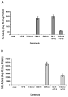
The bidirectional system mediates simultaneous expression of both fl (A) and hrl (B) genes. N2a cells were transiently transfected with water (mock), activator plasmid alone (VP16), reporter plasmid alone (fl-G5-hrl), CMV-fl, CMV-hrl, Unidirectional fl TSTA system (G5fl+VP16), or both reporter and activator (fl-G5-hrl+VP16). The cells were harvested 24 hr after transfection and assayed for FL and hRL activity. Error bars represent the SEM for triplicate measurements.
FIG. 3
FIG. 3
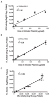
Linear increase in gene expression with increasing doses of the activator plasmid. (A) and (B) show the linear increase in expression of the hrl and fl genes, respectively, as the activator plasmid (VP16) dose is increased from 0 to 0.75 μg/well while the dose of reporter plasmid is maintained at 0.5 μg/well. Seen here are high correlations (r2 = 0.90 and 0.98 for hrl and fl, respectively) between the dose of activator plasmid used for transfection and protein activities. The results were obtained 24 hr after transfection in N2a cells. Error bars indicate the SEM for triplicate measurements. (C) Correlation between hrl and fl expression in N2a cells transfected with various doses of VP16 and a constant dose of fl-G5-hrl. The correlation is r2 = 0.94.
FIG. 4
FIG. 4
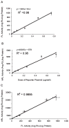
Linear increase in gene expression with increasing levels of reporter plasmid. (A) and (B) show the linear relationship between the expression of fl (A) and hrl (B) genes and the dose of the reporter plasmid (fl-G5-hrl) (0–0.75 μg/well) while the dose of the activator plasmid was maintained at 0.5 μg/well. Seen here are high correlations (r2 = 0.96 and 0.98 for hrl and fl, respectively) between the dose of reporter plasmid used for transfection and protein activities. The results were obtained 24 hr after transfection in N2a cells. Error bars indicate the SEM for triplicate measurements. (C) Correlation between hrl and fl expression in N2a cells transfected with various doses of fl-G5-hrl and a constant dose of VP16. The correlation is r2 = 0.99.
FIG. 5
FIG. 5
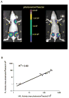
(A) Optical CCD imaging of a mouse carrying N2a cells transiently cotransfected with a constant dose of fl-G5-hrl and various doses of the activator plasmid VP16 implanted subcutaneously at four different sites. Sites A–D indicate cells transfected with various doses of VP16 in increasing order. Both images shown are from the same mouse and are formed by superimposing visible light images on the optical CCD bioluminescence image with a scale depicting photons/cm2/sec/steradian (sr). Mice were imaged after tail vein injection of coelenterazine (left) and after intraperitoneal injection of D-luciferin (right). (B) Correlation of fl and hrl gene expression from three mice optically imaged with cells cotransfected with different doses of the activator plasmid (VP16) and a constant dose of the reporter plasmid (fl-G5-hrl). Shown is a plot of hRL activity versus FL activity, expressed as maximum photons/cm2/sec/sr, obtained from ROIs drawn over the implantation sites on the mouse images (r2 = 0.82, n = 3 mice).
FIG. 6
FIG. 6
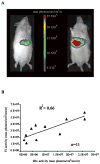
(A) Optical CCD imaging of a mouse injected hydrodynamically with 10 μg each of activator (VP16) and reporter plasmid (fl-G5-hrl) 24 hr before imaging. Both images shown are from the same mouse and are formed by superimposing visible light images on the optical CCD bioluminescence image with a scale depicting photons/cm2/sec/sr. Mice were imaged after tail vein injection of coelenterazine (40 μg; left) and after intraperitoneal injection of D-luciferin (3 mg; right). (B) Correlation of fl and hrl gene expression from mice optically imaged after the injection of 10 μg each of the activator plasmid (VP16) and the reporter plasmid (fl-G5-hrl). Shown is a plot of hRL activity versus FL activity, expressed as maximum photons/cm2/sec/sr, obtained from same-sized ROIs drawn over the liver region on the mouse images (r2 = 0.66, n = 11 mice).

Similar articles

Cited by

References

    1. BARON U, FREUNDLIEB S, GOSSEN M, BUJARD H. Coregulation of two gene activities by tetracycline via a bidirectional promoter. Nucleic Acids Res. 1995;23:3605–3606. - PMC - PubMed
    1. BHAUMIK S, GAMBHIR SS. Optical imaging of Renilla luciferase reporter gene expression in living mice. Proc Natl Acad Sci USA. 2002a;99:377–382. - PMC - PubMed
    1. BHAUMIK S, GAMBHIR SS. Simultaneous imaging of the expression of two bioluminescent reporter genes in living mice. Mol Ther. 2002b;5:S422.
    1. BLAKE P, JOHNSON B, VANMETER JW. Positron emission tomography (PET) and single photon emission computed tomography (SPECT): Clinical applications. J Neuroophthalmol. 2003;23:34–41. - PubMed
    1. CHAPPELL SA, EDELMAN GM, MAURO VP. A 9-nt segment of a cellular mRNA can function as an internal ribosome entry site (IRES) and when present in linked multiple copies greatly enhances IRES activity. Proc Natl Acad Sci USA. 2000;97:1536–1541. - PMC - PubMed

Publication types