

doi: 10.1016/j.str.2004.04.024.
West Nile virus core protein; tetramer structure and ribbon formation
Affiliations
- PMID: 15242592
- PMCID: PMC7173237
- DOI: 10.1016/j.str.2004.04.024
Item in Clipboard
West Nile virus core protein; tetramer structure and ribbon formation
Structure.
2004 Jul.
Abstract
We have determined the crystal structure of the core (C) protein from the Kunjin subtype of West Nile virus (WNV), closely related to the NY99 strain of WNV, currently a major health threat in the U.S. WNV is a member of the Flaviviridae family of enveloped RNA viruses that contains many important human pathogens. The C protein is associated with the RNA genome and forms the internal core which is surrounded by the envelope in the virion. The C protein structure contains four alpha helices and forms dimers that are organized into tetramers. The tetramers form extended filamentous ribbons resembling the stacked alpha helices seen in HEAT protein structures.
Figures

Sequence Alignment of the C Proteins of Kunjin Virus, the NY99 and Nigerian Strains of WNV, and Dengue The start and end of the trypsin-cleaved fragment (bullets) as well as the site of maturation cleavage (arrowhead) are indicated. The position of the α helices in the C structure are shown above the sequence (spirals) (Gouet et al., 1999).

Stereoviews of the WNV C Protein Structure (A) Detail of the weighted 2Fo − Fc electron density map, showing part of helix α4 from Asn72 to Ile94 (Esnouf, 1999). (B) Cα backbone trace of CD dimer. C, blue; D, red. (C) Superposition of subunit A (blue) with residues B1108 to B1206 of Pyrococcus woesei TFB (PDB code 1AIS; green), aligned using the helical segments identified by the DALI search (Holm and Sander, 1995). (D) Superposition of the C protein CD dimer (purple) with the solution structure dimer of dengue C protein (1R6R) (green). The least mean square fit was calculated using residues 54–96.
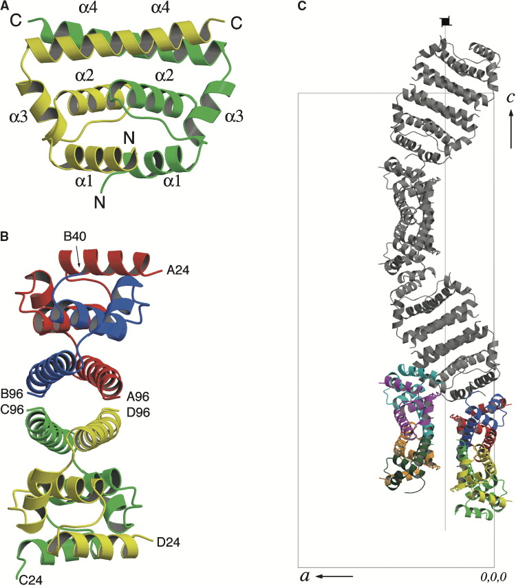
Ribbon Diagrams of the WNV C Protein The diagrams have been colored as follows: A, red; B, blue; C, green; D, yellow; E, cyan; F, magenta; G, orange; H, teal. (A) Ribbon diagram of CD dimer (Esnouf, 1999). Secondary structure elements are indicated. (B) Ribbon diagram of ABDC tetramer looking down the tunnel. N- and C-terminal residues are indicated. (C) Arrangement of tetramers in the crystal, viewed down the b axis. The unit cell, the crystal a and c axes and the 4-fold screw axis are indicated. The ABCD and EFGH tetramers in the asymmetric unit are shown in color, while the symmetry-related ABCD tetramers that form one half of the tubule are shown in gray. The symmetry-related EFGH tetramers are omitted for clarity.

Interactions and Surface Potential of the WNV Core Protein Tetramer (A) View of the interface between the AB dimer (red, blue) and the symmetry-related C′D′ dimer (green, yellow) (Esnouf, 1999). The position that the missing helix α1 in the B subunit would have had where it overlaps with subunit C′ is shown in gray. The Tyr and Leu residues that comprise the hydrophobic pocket in AB are indicated, as is the density enclosed by the pocket in AB and C′D′. (B) Electrostatic potential surface of C protein tetramer, viewed in the same orientation as Figure 3B.
Comment in
-
Visualizing the house from the brick.Structure. 2004 Jul;12(7):1119-20. doi: 10.1016/j.str.2004.06.009. Structure. 2004. PMID: 15242583 No abstract available.
References
-
- Andrade M.A., Petosa C., O'Donoghue S.I., Müller C.W., Bork P. Comparison of ARM and HEAT protein repeats. J. Mol. Biol. 2001;309:1–18. - PubMed
-
- Brinton M.A. The molecular biology of West Nile virus: a new invader of the western hemisphere. Annu. Rev. Microbiol. 2002;56:371–402. - PubMed
-
- Buckley A., Dawson A., Moss S.R., Hinsley S.A., Bellamy P.E., Gould E.A. Serological evidence of West Nile virus, Usuru virus and Sindbis virus infection of birds in the UK. J. Gen. Virol. 2003;84:2807–2817. - PubMed
-
- CDC. (2004). West Nile Virus: Statistics, Surveillance and Control (Center for Disease Control). http://www.cdc.gov/ncidod/dvbid/westnile/surv&control.htm.
-
- Choi H.-K., Tong L., Minor W., Dumas P., Boege U., Rossmann M.G., Wengler G. Structure of Sindbis virus core protein reveals a chymotrypsin-like serine protease and the organization of the virion. Nature. 1991;354:37–43. - PubMed
MeSH terms
Substances
Associated data
- Actions
LinkOut - more resources
Full Text Sources
Other Literature Sources
