

Antigenic evidence of prevalence and diversity of Mycobacterium tuberculosis arabinomannan
- PMID: 15243086
- PMCID: PMC446310
- DOI: 10.1128/JCM.42.7.3225-3231.2004
Antigenic evidence of prevalence and diversity of Mycobacterium tuberculosis arabinomannan
Abstract
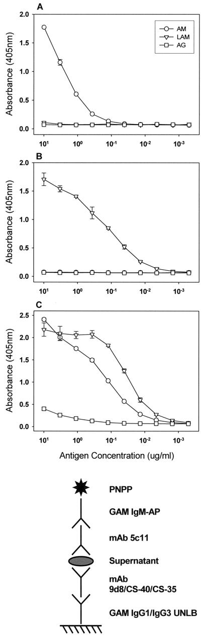
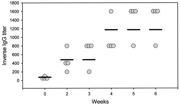
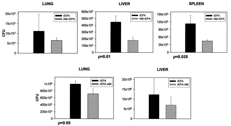
Arabinomannan (AM) is a polysaccharide of the mycobacterial capsule. The capsular polysaccharides of various microorganisms are diverse, and this diversity is important for classification of organisms into serotypes and vaccine development. In the present study we examined the prevalence and diversity of AM among Mycobacterium tuberculosis strains using four AM-binding monoclonal antibodies (MAbs). One of these MAbs, MAb 9d8, is known to bind to AM specifically. By whole-cell enzyme-linked immunosorbent assay (ELISA), the AM recognized by MAb 9d8 was detected on the surfaces of 9 of 11 strains, while 2 strains showed no reactivity with MAb 9d8. However, the AM recognized by MAb 9d8 was found in the culture supernatants of all 11 M. tuberculosis strains tested, as demonstrated by capture ELISA. Other AM-binding MAbs reacted both with the surfaces and with the culture supernatants of all 11 strains. Mice immunized with an experimental AM-recombinant Pseudomonas aeruginosa exoprotein A (rEPA) conjugate vaccine had an increased antibody response to AM and a moderate reduction in the numbers of CFU in their organs 7 days after challenge. Our results indicate that AM was detected in all M. tuberculosis strains tested, with differences in epitope distributions of certain strains. In addition, our results suggest that an experimental AM-rEPA vaccine has a moderate effect on the numbers of CFU in organs early after infection.
Figures
References
-
- Besra, G. S., and D. Chatterjee. 1994. Lipids and carbohydrates of Mycobacterium tuberculosis, p. 285-306. In B. R. Bloom (ed.), Tuberculosis: pathogenesis, protection, and control. ASM Press, Washington, D.C.
-
- Centers for Disease Control and Prevention. 1996. The role of BCG vaccine in the prevention and control of tuberculosis in the United States. A joint statement by the Advisory Counsil for the Elimination of Tuberculosis and the Advisory Committee on Immunization Practices. Morb. Mortal. Wkly. Rep. 45:1-18. - PubMed
-
- Chatterjee, D., K. Lowell, B. Rivoire, M. R. McNeil, and P. J. Brennan. 1992. Lipoarabinomannan of Mycobacterium tuberculosis. Capping with mannosyl residues in some strains. J. Biol. Chem. 267:6234-6239. - PubMed
-
- Collins, H. L., and S. H. E. Kaufmann. 2001. Prospects for better tuberculosis vaccines. Lancet Infect. Dis. 1:21-28. - PubMed
-
- Daffe, M., and P. Draper. 1998. The envelope layers of mycobacteria with reference to their pathogenicity. Adv. Microb. Physiol. 39:131-203. - PubMed
Publication types
MeSH terms
Substances
Grants and funding
- R21 AI043268/AI/NIAID NIH HHS/United States
- AI053192/AI/NIAID NIH HHS/United States
- R01 AI033142/AI/NIAID NIH HHS/United States
- R01 AI033774/AI/NIAID NIH HHS/United States
- R37 AI026170/AI/NIAID NIH HHS/United States
- R01 HL059842/HL/NHLBI NIH HHS/United States
- AI033142/AI/NIAID NIH HHS/United States
- AI033774/AI/NIAID NIH HHS/United States
- R03 AI053192/AI/NIAID NIH HHS/United States
- HL059842/HL/NHLBI NIH HHS/United States
- R01 AI043268/AI/NIAID NIH HHS/United States
- AI026170/AI/NIAID NIH HHS/United States
- R01 AI052733/AI/NIAID NIH HHS/United States
- AI052733/AI/NIAID NIH HHS/United States
- R01 AI026170/AI/NIAID NIH HHS/United States
- K08 AI001691/AI/NIAID NIH HHS/United States
- R37 AI033142/AI/NIAID NIH HHS/United States
- AI043268/AI/NIAID NIH HHS/United States
- AI001691/AI/NIAID NIH HHS/United States
LinkOut - more resources
Full Text Sources
Other Literature Sources
