Is a single anterolateral screw-plate fixation sufficient for the treatment of spinal fractures in the thoracolumbar junction? A biomechanical in vitro investigation
- PMID: 15243790
- PMCID: PMC3476694
- DOI: 10.1007/s00586-004-0770-9
Is a single anterolateral screw-plate fixation sufficient for the treatment of spinal fractures in the thoracolumbar junction? A biomechanical in vitro investigation
Abstract
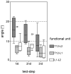
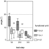
Controversy exists about the indications, advantages and disadvantages of various surgical techniques used for anterior interbody fusion of spinal fractures in the thoracolumbar junction. The purpose of this study was to evaluate the stabilizing effect of an anterolateral and thoracoscopically implantable screw-plate system. Six human bisegmental spinal units (T12-L2) were used for the biomechanical in vitro testing procedure. Each specimen was tested in three different scenarios: (1) intact spinal segments vs (2) monosegmental (T12/L1) anterolateral fixation (macsTL, Aesculap, Germany) with an interbody bone strut graft from the iliac crest after both partial corpectomy (L1) and discectomy (T12/L1) vs (3) bisegmental anterolateral instrumentation after extended partial corpectomy (L1), and bisegmental discectomy (T12/L1 and L1/L2). Specimens were loaded with an alternating, nondestructive maximum bending moment of +/-7.5 Nm in six directions: flexion/extension, right and left lateral bending, and right and left axial rotation. Motion analysis was performed by a contact-less three-dimensional optical measuring system. Segmental stiffness of the three different scenarios was evaluated by the relative alteration of the intervertebral angles in the three main anatomical planes. With each stabilization technique, the specimens were more rigid, compared with the intact spine, for flexion/extension (sagittal plane) as well as in left and right lateral bending (frontal plane). In these planes the bisegmental instrumentation compared to the monosegmental case had an even larger stiffening effect on the specimens. In contrast to these findings, axial rotation showed a modest increase of motion after bisegmental instrumentation. To conclude, the immobilization of monosegmental fractures in the thoracolumbar junction can be secured by means of bone grafting and the implant used in this study for all three anatomical planes. After bisegmental anterolateral stabilization a sufficient reduction of the movements was registered for flexion/extension and lateral bending. However, the observed slight increase of the range of motion in the transversal plane may lead to loosening of the implant before union. Therefore, the use of an additional dorsal fixation device should be considered.
Figures


Similar articles
-
Two column lesions in the thoracolumbar junction: anterior, posterior or combined approach? A comparative biomechanical in vitro investigation.Eur Spine J. 2007 Jun;16(6):813-20. doi: 10.1007/s00586-006-0201-1. Epub 2006 Aug 30. Eur Spine J. 2007. PMID: 16944226 Free PMC article.
-
Biomechanical evaluation of anterior thoracolumbar spinal instrumentation.Spine (Phila Pa 1976). 1995 Sep 15;20(18):1979-83. doi: 10.1097/00007632-199509150-00003. Spine (Phila Pa 1976). 1995. PMID: 8578371
-
Non-fusion instrumentation of the lumbar spine with a hinged pedicle screw rod system: an in vitro experiment.Eur Spine J. 2009 Oct;18(10):1478-85. doi: 10.1007/s00586-009-1052-3. Epub 2009 Jun 6. Eur Spine J. 2009. PMID: 19504129 Free PMC article.
-
Biomechanical evaluation of lateral lumbar interbody fusion with secondary augmentation.J Neurosurg Spine. 2016 Dec;25(6):720-726. doi: 10.3171/2016.4.SPINE151386. Epub 2016 Jul 8. J Neurosurg Spine. 2016. PMID: 27391398
-
[MAIGNE'S THORACOLUMBAR JUNCTION SYNDROME].Harefuah. 2024 Dec;163(11):728-731. Harefuah. 2024. PMID: 39692376 Review. Hebrew.
Cited by
-
En bloc spondylectomy reconstructions in a biomechanical in-vitro study.Eur Spine J. 2008 May;17(5):715-25. doi: 10.1007/s00586-008-0588-y. Epub 2008 Jan 15. Eur Spine J. 2008. PMID: 18196295 Free PMC article.
-
Contribution of Round vs. Rectangular Expandable Cage Endcaps to Spinal Stability in a Cadaveric Corpectomy Model.Int J Spine Surg. 2015 Oct 22;9:53. doi: 10.14444/2053. eCollection 2015. Int J Spine Surg. 2015. PMID: 26609508 Free PMC article.
-
Stepwise resection of the posterior ligamentous complex for stability of a thoracolumbar compression fracture: An in vitro biomechanical investigation.Medicine (Baltimore). 2017 Sep;96(35):e7873. doi: 10.1097/MD.0000000000007873. Medicine (Baltimore). 2017. PMID: 28858098 Free PMC article.
-
Two column lesions in the thoracolumbar junction: anterior, posterior or combined approach? A comparative biomechanical in vitro investigation.Eur Spine J. 2007 Jun;16(6):813-20. doi: 10.1007/s00586-006-0201-1. Epub 2006 Aug 30. Eur Spine J. 2007. PMID: 16944226 Free PMC article.
-
The Effect of Polymethyl Methacrylate Augmentation on the Primary Stability of Cannulated Bone Screws in an Anterolateral Plate in Osteoporotic Vertebrae: A Human Cadaver Study.Global Spine J. 2016 Feb;6(1):46-52. doi: 10.1055/s-0035-1555659. Epub 2015 Jun 15. Global Spine J. 2016. PMID: 26835201 Free PMC article.
References
-
- An Spine. 1995;20:1979. - PubMed
-
- Been Acta. 1999;Neurochir:349. - PubMed
-
- Beisse Eur J Trauma. 2001;27:275.
-
- Beisse R, Potulski M, Beger J, Bühren V (2002) Development and clinical application of a thoracoscopically implantable anterior plate for treatment of thoracolumbar fractures and instabilities. Orthopäde 31:413–422 - PubMed
-
- Bernhardt Spine. 1989;14:717. - PubMed
MeSH terms
LinkOut - more resources
Full Text Sources
Medical

