

High prevalence of cutaneous human papillomavirus DNA on the top of skin tumors but not in "Stripped" biopsies from the same tumors
- PMID: 15245440
- PMCID: PMC3822504
- DOI: 10.1111/j.0022-202X.2004.23205.x
High prevalence of cutaneous human papillomavirus DNA on the top of skin tumors but not in "Stripped" biopsies from the same tumors
Abstract
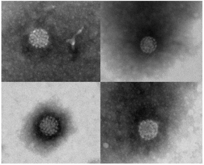
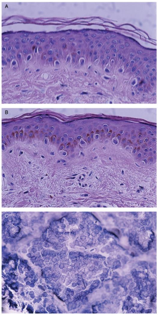
Genomes of human papillomaviruses (HPV) are common in biopsies from non-melanoma skin cancers but are also found on healthy skin and it is possible that HPV positivity in tumor biopsies by PCR may merely reflect contamination of the lesion surface. To investigate this issue, 229 immunocompetent patients were tested for HPV DNA in swab samples collected on top of skin tumors and in biopsies of the same tumors, obtained after stripping with tape to remove superficial layers. HPV DNA was detected on top of 69% (159 of 229) of the lesions, and in 12% (28 of 229) of the stripped biopsies (p<0.001). The difference was seen for all four types of tumors studied. Seborrheic keratosis had 79% (34 of 43) HPV positivity on top of lesions versus 19% (eight of 43) in biopsies; actinic keratosis had 83% (38 of 46) HPV positivity on top versus 11% (five of 46) in biopsies; basal cell carcinoma had 63% (69 of 109) on top versus 8% (nine of 109) in biopsies and squamous cell carcinoma had 58% (18 of 31) on top versus 19% (six of 31) in biopsies. HPV DNA is common in superficial layers of lesions, but is not necessarily present throughout tumors.
Figures
Similar articles
-
Four novel human betapapillomaviruses of species 2 preferentially found in actinic keratosis.J Gen Virol. 2008 Oct;89(Pt 10):2467-2474. doi: 10.1099/vir.0.2008/001925-0. J Gen Virol. 2008. PMID: 18796715
-
Viral DNA detection and RAS mutations in actinic keratosis and nonmelanoma skin cancers.Br J Dermatol. 2010 Feb 1;162(2):325-31. doi: 10.1111/j.1365-2133.2009.09480.x. Epub 2009 Aug 29. Br J Dermatol. 2010. PMID: 19849697
-
Pattern of HPV infection in basal cell carcinoma and in perilesional skin biopsies from immunocompetent patients.Virol J. 2012 Dec 17;9:309. doi: 10.1186/1743-422X-9-309. Virol J. 2012. PMID: 23244448 Free PMC article.
-
Human papillomavirus infection and skin cancer risk in organ transplant recipients.J Investig Dermatol Symp Proc. 2001 Dec;6(3):207-11. doi: 10.1046/j.0022-202x.2001.00048.x. J Investig Dermatol Symp Proc. 2001. PMID: 11924829 Review.
-
HPV in oral squamous cell carcinoma vs head and neck squamous cell carcinoma biopsies: a meta-analysis (1988-2007).Ann Oncol. 2008 Oct;19(10):1681-90. doi: 10.1093/annonc/mdn372. Epub 2008 Jun 16. Ann Oncol. 2008. PMID: 18558666 Review.
Cited by
-
The Role of Beta HPV Types and HPV-Associated Inflammatory Processes in Cutaneous Squamous Cell Carcinoma.J Immunol Res. 2020 Apr 6;2020:5701639. doi: 10.1155/2020/5701639. eCollection 2020. J Immunol Res. 2020. PMID: 32322596 Free PMC article. Review.
-
De novo sequence assembly requires bioinformatic checking of chimeric sequences.PLoS One. 2020 Aug 10;15(8):e0237455. doi: 10.1371/journal.pone.0237455. eCollection 2020. PLoS One. 2020. PMID: 32777809 Free PMC article.
-
Relationship of human papillomavirus with seborrheic keratosis of the female genital tract - a case-series and literature review.Histol Histopathol. 2021 Dec;36(12):1209-1218. doi: 10.14670/HH-18-357. Epub 2021 May 25. Histol Histopathol. 2021. PMID: 34170001 Review.
-
The oral cavity contains abundant known and novel human papillomaviruses from the Betapapillomavirus and Gammapapillomavirus genera.J Infect Dis. 2011 Sep 1;204(5):787-92. doi: 10.1093/infdis/jir383. J Infect Dis. 2011. PMID: 21844305 Free PMC article.
-
Cutaneous human papillomaviruses found in sun-exposed skin: Beta-papillomavirus species 2 predominates in squamous cell carcinoma.J Infect Dis. 2007 Sep 15;196(6):876-83. doi: 10.1086/521031. Epub 2007 Aug 6. J Infect Dis. 2007. PMID: 17703418 Free PMC article.
References
-
- Antonsson A, Erfurt C, Hazard H, et al. Prevalence and type spectrum of human papillomaviruses in healthy skin samples collected in three continents. J Gen Virol. 2003a;84:1881–1886. - PubMed
-
- Astori G, Lavergne D, Benton C, Hockmayr B, Egawa K, Garbe C, de Villiers EM. Human papillomaviruses are commonly found in normal skin of immunocompetent hosts. J Invest Dermatol. 1998;110:752–755. - PubMed
-
- Bens G, Wieland U, Hofmann A, Hopfl R, Pfister H. Detection of new human papillomavirus sequences in skin lesions of a renal transplant recipient and characterization of one complete genome related to epidermodysplasia verruciformis-associated types. J Gen Virol. 1998;79:779–787. - PubMed
Publication types
MeSH terms
Substances
Grants and funding
LinkOut - more resources
Full Text Sources
Other Literature Sources
Medical
