

A highly pathogenic porcine reproductive and respiratory syndrome virus generated from an infectious cDNA clone retains the in vivo virulence and transmissibility properties of the parental virus
- PMID: 15246270
- PMCID: PMC7127741
- DOI: 10.1016/j.virol.2004.04.046
A highly pathogenic porcine reproductive and respiratory syndrome virus generated from an infectious cDNA clone retains the in vivo virulence and transmissibility properties of the parental virus
Abstract
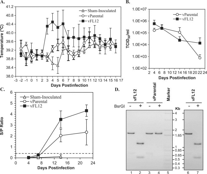
The nucleotide sequence of a highly pathogenic porcine reproductive and respiratory syndrome virus (PRRSV) was determined. Transfection of MARC-145 cells with capped in vitro transcripts derived from a full-length cDNA clone of the viral genome resulted in infectious PRRSV with growth characteristics similar to that of the parental virus. Primer extension analysis revealed that during replication, the viral polymerase corrected the two nonviral guanosine residues present at the 5' terminus of the transfected transcripts. Animal studies showed that the cloned virus induced hyperthermia, persistent viremia, and antibody response, similar to that observed with the parental virus. Contact transmission occurred rapidly within 3 days of introduction of naïve pigs into the group of clone virus-inoculated pigs. These results suggest that the cloned virus retains the in vivo virulence and contagion properties of the parental virus, thus, providing the background for reverse genetics manipulation in systematic examination of attenuation and virulence phenotypes.
Figures
Similar articles
-
Generation of an infectious clone of VR-2332, a highly virulent North American-type isolate of porcine reproductive and respiratory syndrome virus.J Virol. 2003 Mar;77(6):3702-11. doi: 10.1128/jvi.77.6.3702-3711.2003. J Virol. 2003. PMID: 12610145 Free PMC article.
-
The 30-amino-acid deletion in the Nsp2 of highly pathogenic porcine reproductive and respiratory syndrome virus emerging in China is not related to its virulence.J Virol. 2009 May;83(10):5156-67. doi: 10.1128/JVI.02678-08. Epub 2009 Feb 25. J Virol. 2009. PMID: 19244318 Free PMC article.
-
Direct inoculation of RNA transcripts from an infectious cDNA clone of porcine reproductive and respiratory syndrome virus (PRRSV) into the lymph nodes and tonsils of pigs initiates PRRSV infection in vivo.Arch Virol. 2007;152(7):1383-7. doi: 10.1007/s00705-007-0955-8. Epub 2007 Mar 15. Arch Virol. 2007. PMID: 17361326
-
[Advances in Understanding of the Infection/Replication Mechanisms and Virulence Determinants of the Porcine Reproductive and Respiratory Syndrome Virus].Bing Du Xue Bao. 2015 Sep;31(5):585-92. Bing Du Xue Bao. 2015. PMID: 26738300 Review. Chinese.
-
Engineering the PRRS virus genome: updates and perspectives.Vet Microbiol. 2014 Dec 5;174(3-4):279-295. doi: 10.1016/j.vetmic.2014.10.007. Epub 2014 Oct 23. Vet Microbiol. 2014. PMID: 25458419 Free PMC article. Review.
Cited by
-
Harnessing longitudinal information to identify genetic variation in tolerance of pigs to Porcine Reproductive and Respiratory Syndrome virus infection.Genet Sel Evol. 2018 Oct 24;50(1):50. doi: 10.1186/s12711-018-0420-z. Genet Sel Evol. 2018. PMID: 30355341 Free PMC article.
-
A Synthetic Porcine Reproductive and Respiratory Syndrome Virus Strain Confers Unprecedented Levels of Heterologous Protection.J Virol. 2015 Dec;89(23):12070-83. doi: 10.1128/JVI.01657-15. Epub 2015 Sep 23. J Virol. 2015. PMID: 26401031 Free PMC article.
-
Immune evasion of porcine reproductive and respiratory syndrome virus through glycan shielding involves both glycoprotein 5 as well as glycoprotein 3.J Virol. 2011 Jun;85(11):5555-64. doi: 10.1128/JVI.00189-11. Epub 2011 Mar 16. J Virol. 2011. PMID: 21411530 Free PMC article.
-
Genetic relationships of antibody response, viremia level, and weight gain in pigs experimentally infected with porcine reproductive and respiratory syndrome virus1.J Anim Sci. 2018 Sep 7;96(9):3565-3581. doi: 10.1093/jas/sky229. J Anim Sci. 2018. PMID: 29905795 Free PMC article.
-
Comparison of host genetic factors influencing pig response to infection with two North American isolates of porcine reproductive and respiratory syndrome virus.Genet Sel Evol. 2016 Jun 20;48(1):43. doi: 10.1186/s12711-016-0222-0. Genet Sel Evol. 2016. PMID: 27324857 Free PMC article.
References
-
- Albina E. Epidemiology of porcine reproductive and respiratory syndrome (PRRS): an overview. Vet. Microbiol. 1997;55:309–316. - PubMed
-
- Andrejeva J., Puurand U., Merits A., Rabenstein F., Jarvekulg L., Valkonen J.P. Potyvirus helper component-proteinase and coat protein (CP) have coordinated functions in virus–host interactions and the same CP motif affects virus transmission and accumulation. J. Gen. Virol. 1999;80(Pt 5):1133–1139. - PubMed
-
- Bastos R.G., Dellagostin O.A., Barletta R.G., Doster A.R., Nelson E., Zuckermann F., Osorio F.A. Immune response of pigs inoculated with Mycobacterium bovis BCG expressing a truncated form of GP5 and M protein of porcine reproductive and respiratory syndrome virus. Vaccine. 2004;22:467–474. - PubMed
-
- Boyer J.C., Haenni A.L. Infectious transcripts and cDNA clones of RNA viruses. Virology. 1994;198:415–426. - PubMed
Publication types
MeSH terms
Substances
Grants and funding
LinkOut - more resources
Full Text Sources
Other Literature Sources
